. 2019 Jun 7;124(12):1747-1759.
doi: 10.1161/CIRCRESAHA.119.314845. Epub 2019 Apr 22.

Complement Membrane Attack Complexes Assemble NLRP3 Inflammasomes Triggering IL-1 Activation of IFN-γ-Primed Human Endothelium

Affiliations

Complement Membrane Attack Complexes Assemble NLRP3 Inflammasomes Triggering IL-1 Activation of IFN-γ-Primed Human Endothelium

Catherine B Xie et al. Circ Res. .

Erratum in

Abstract

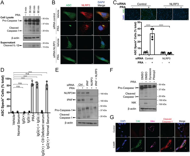
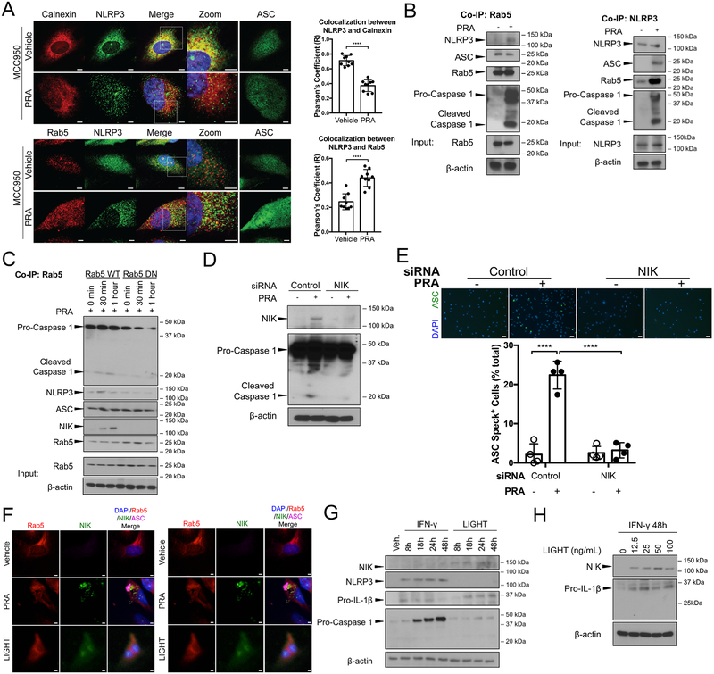
Rationale: Complement activation contributes to multiple immune-mediated pathologies. In late allograft failure, donor-specific antibody deposits complement membrane attack complexes (MAC) on graft endothelial cells (ECs), substantially increasing their immunogenicity without causing lysis. Internalized MAC stabilize NIK (NF-κB [nuclear factor kappa-light-chain-enhancer of activated B cells]-inducing kinase) protein on Rab5+MAC+ endosomes, activating noncanonical NF-κB signaling. However, the link to increased immunogenicity is unclear.

Objective: To identify mechanisms by which alloantibody and internalized MAC activate ECs to enhance their ability to increase T-cell responses.

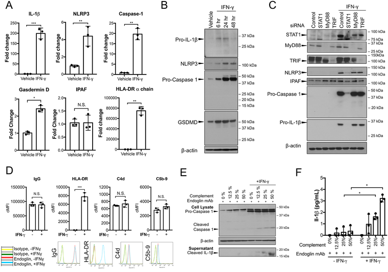
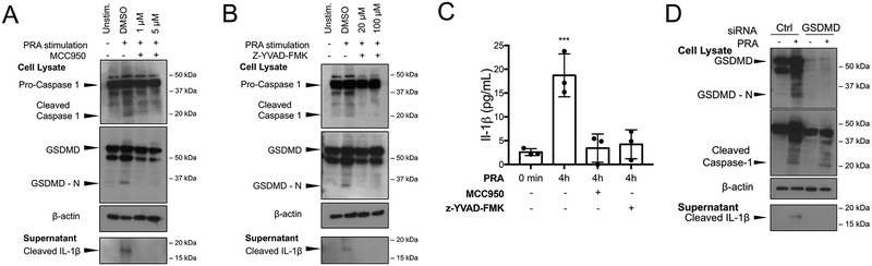
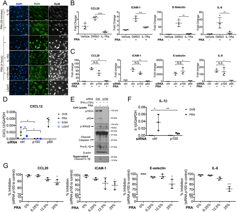
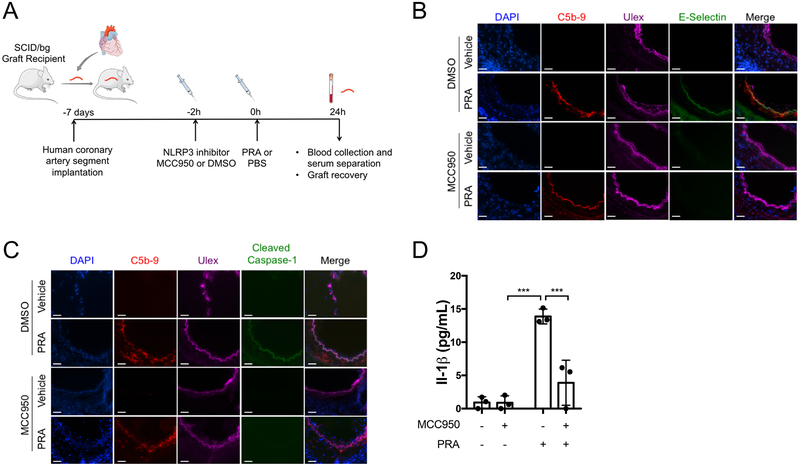
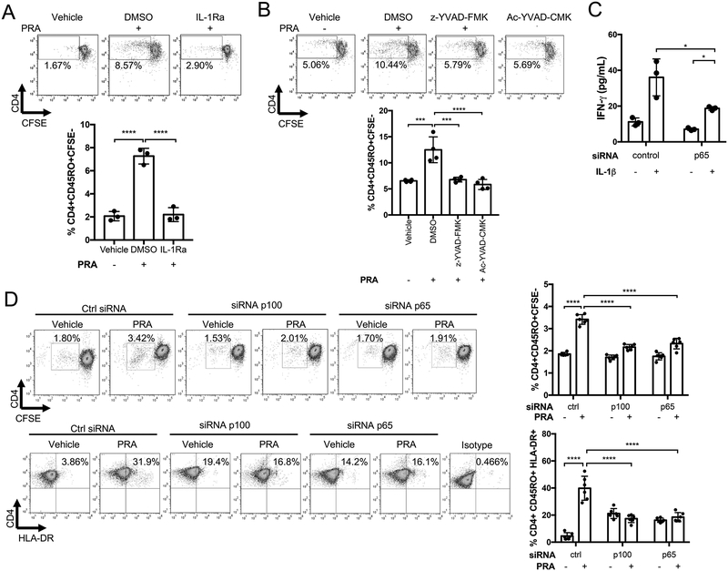
Methods and results: In human EC cultures, internalized MAC also causes NLRP3 (NOD-like receptor family pyrin domain containing 3) translocation from endoplasmic reticulum to Rab5+MAC+NIK+ endosomes followed by endosomal NIK-dependent inflammasome assembly. Cytosolic NIK, stabilized by LIGHT (lymphotoxin-like inducible protein that competes with glycoprotein D for herpesvirus entry on T cells), does not trigger inflammasome assembly, and ATP-triggered inflammasome assembly does not require NIK. IFN-γ (interferon-γ) primes EC responsiveness to MAC by increasing NLRP3, pro-caspase 1, and gasdermin D expression. NIK-activated noncanonical NF-κB signaling induces pro-IL (interleukin)-1β expression. Inflammasome processed pro-IL-1β, and gasdermin D results in IL-1β secretion that increases EC immunogenicity through IL-1 receptor signaling. Activation of human ECs lining human coronary artery grafts in immunodeficient mouse hosts by alloantibody and complement similarly depends on assembly of an NLRP3 inflammasome. Finally, in renal allograft biopsies showing chronic rejection, caspase-1 is activated in C4d+ ECs of interstitial microvessels, supporting the relevance of the cell culture findings.

Conclusions: In response to antibody-mediated complement activation, IFN-γ-primed human ECs internalize MAC, triggering both endosomal-associated NIK-dependent NLRP3 inflammasome assembly and IL-1 synthesis, resulting in autocrine/paracrine IL-1β-mediated increases in EC immunogenicity. Similar responses may underlie other complement-mediated pathologies.

Keywords: endosomes; endothelium; immune system; inflammasomes; transplantation.


Figures

Figure 1.
Figure 1.. Alloantibody-mediated MAC deposition on IFN-γ-pretreated human ECs activates an NLRP3 inflammasome.
(A) ECs pre-treated with IFN-γ (50ng/mL) for 48 hours prior to PRA sera treatment for the indicated times and whole cell lysates and culture supernatants were assessed for cleaved caspase-1 and cleaved IL-1β, respectively, by immunoblot. (B) ASC-GFP transduced and IFN-γ pre-treated ECs were transfected with control or NLRP3 siRNA and treated with PRA sera prior to confocal microscopy staining for NLRP3. Scale bar, 2 μm. (C) Top: ASC speck formation of control and NLRP3 siRNA transfected PRA-treated ASC-GFP ECs were assessed by epifluorescence. Scale bar, 50 μm. Bottom: Each symbol represents the mean of the % ASC speck+ cells in 3 low powered fields per sample (n=4, one-way ANOVA and Tukey’s multiple comparisons test, SEM). (D) PRA sera was separated into IgG(+) and IgG(−) fractions and added to IFN-γ pre-treated ASC-GFP ECs either alone, or in combination with C9 deficient or normal serum as indicated for ASC speck formation analysis. Each symbol represents the mean of the % ASC speck+ cells in at least 3 low powered fields per sample. (n=3, one-way ANOVA and Tukey’s multiple comparisons test, SEM). (E) IFN-γ pre-treated ECs were transfected with either control, IPAF, NLRP3 or both IPAF and NLRP3 siRNA prior to treatment with PRA and cell lysates were assessed for cleaved caspase-1 by immunoblot. (F) IFN-γ-primed ECs were pre-treated with dynasore or pitstop2, two pharmacological inhibitors of clathrin-mediated endocytosis, treated with PRA sera for 30 minutes. Whole cell lysates were analyzed for cleaved caspase-1 and NIK by immunoblot. (G) Renal biopsies from patients with chronic antibody mediated rejection (CAMR) or taken at the time of transplant (control) were co-stained for C4d, Ulex and cleaved caspase-1 and analyzed by confocal microscopy. Scale bar, 5 μm. ****P<0.0001, n.s., non-significant.
Figure 2.
Figure 2.. IFN-γ primes human ECs for MAC-induced NLRP3 inflammasome activation.
(A) ECs were primed with IFN-γ (50ng/mL) for 24 hours and upregulation of IL-1β, NLRP3, NLRC4/IPAF, Caspase-1, Gasdermin D and HLA-DR α-chain transcript levels were assessed by qRT-PCR (n=3, Student’s t-test, SEM). (B) ECs were stimulated with IFN-γ for the duration indicated and pro-IL-1β, NLRP3, pro-caspase-1, and gasdermin D expression were assessed by immunoblot. (C) Unprimed and IFN-γ-primed ECs were transfected with control, STAT1, MyD88, or TRIF siRNA and protein expression of NLRP3, IPAF, pro-caspase-1 and pro-IL-1β were assessed by immunoblot. (D) Unprimed and IFN-γ-primed ECs were incubated with complement-fixing anti-human endoglin IgG2a mouse monoclonal antibody (mAb) (20μg/mL) and human complement prior to flow cytometry analysis of IgG binding, HLA-DR, C4d and C5b-9 (n=3, Student’s t-test, SEM). (E) Unprimed and IFN-γ-primed ECs were incubated with anti-human endoglin IgG2a mAb and varying concentrations of human complement. Cell lysates and culture supernatants were assessed for cleaved caspase-1 and cleaved IL-1β, respectively, by immunoblot. (F) ELISA measurement of IL-1β secretion in culture supernatants by unprimed and IFN-γ-primed ECs after incubation with anti-human endoglin mAb and varying levels of human complement (n=3, one-way ANOVA and Tukey’s multiple comparisons test, SEM). *P<0.05, **P<0.01, ***P<0.001, n.s., non-significant.
Figure 3.
Figure 3.. Dual role of Rab5+ endosome-associated NIK in non-canonical NF-κB activation and inflammasome assembly for sustained pro-IL-1β expression and processing, respectively.
(A) Left: Confocal imaging analysis of IFN-γ-primed ASC-GFP ECs that were pretreated with NLRP3 inhibitor MCC950 to prevent ASC oligomerization or control DMSO prior to PRA sera treatment for 30 minutes and co-staining with NLRP3 and Rab5 or calnexin. Scale bar, 5 μm. Right: Pearson’s colocalization coefficients (R) were calculated for calnexin and NLRP3 (top) and Rab5 and NLRP3 (bottom). Each symbol represents a colocalization coefficient R calculated of ECs (n=9, Student t-test, SEM). (B) Rab5 and NLRP3 co-immunoprecipitations (co-IPs) were performed on PRA sera or vehicle treated IFN-γ-primed ECs and immunoblotted as indicated. (C) Stably transduced Rab5-WT or Rab5 DN (S43N) IFN-γ-primed ECs were treated with PRA sera prior to co-immunoprecipitation of Rab5 and subsequent immunoblot analysis for cleaved caspase-1, NLRP3, ASC and NIK. (D) IFN-γ-primed ECs were transfected with control or NIK siRNA and whole cell lysates were analyzed for cleaved caspase-1 and NIK by immunoblot. (E) Top: ASC speck formation in control and NIK siRNA transfected IFN-γ-primed ECs was assessed by epifluorescence. Scale bar, 50 μm. Bottom: Each symbol represents the mean of the percentage of ASC speck+ cells in at least 3 low powered fields per sample (n=4, one-way ANOVA and Tukey’s multiple comparisons test, SEM). (F) Left: ASC-GFP transduced ECs were treated with either PRA sera or LIGHT (100ng/mL) and co-stained for Rab5 and NIK. Scale bar, 5 μm. Right: Confocal imaging analysis of ASC-GFP transduced ECs treated with either PRA sera or LIGHT, prior to RelB staining. Scale bar, 5 μm. (G) ECs were treated with either IFN-γ or LIGHT for 8, 18, 24 or 48 hours and cell lysates were analyzed for NIK, NLRP3, pro-IL-1β, pro-caspase-1 expression by immunoblot. (H) ECs were primed with IFN-γ for 48 hours and subsequently treated with varying concentrations of LIGHT for 18 hours prior to immunoblot analysis of NIK and pro-IL-1β expression. ****P<0.0001.
Figure 4.
Figure 4.. MAC-induced NLRP3 inflammasome induces IL-1β maturation and gasdermin-D-dependent IL-1β release from human ECs.
(A) IFN-γ-primed ECs were pre-treated with NLRP3 inhibitor MCC950 prior to PRA sera treatment and immunoblot analysis of whole cell lysates for cleaved caspase-1 and gasdermin D amino terminal region (GSDMD-N) and culture supernatants for cleaved IL-1β. (B) IFN-γ-primed ECs were pretreated with selective caspase-1 inhibitor z-YVAD-FMK prior to PRA sera treatment and immunoblot analysis of whole cell lysates for cleaved caspase-1 and GSDMD-N and culture supernatants for cleaved IL-1β. (C) ELISA measurements of IL-1β in culture supernatants of IFN-γ-primed ECs pretreated with z-YVAD-FMK or DMSO prior to PRA treatment and compared to IFN-γ-primed ECs simply washed with PRA sera (t=0 control) (n=3, Student t-test, SEM). (D) IFN-γ-primed ECs were transfected with either control or gasdermin D siRNA and whole cell lysates were assessed for cleaved caspase-1 and culture supernatants were assessed for cleaved IL-1β. **P<0.01.
Figure 5.
Figure 5.. EC secreted IL-1 activates autocrine/paracrine induction of pro-inflammatory genes through canonical NF-κB signaling.
(A) IFN-γ-primed ECs were pre-treated with either DMSO, IL-1Ra, caspase-1 inhibitor z-YVAD-FMK prior to PRA sera treatment for 30 minutes or 4 hours, stained for either RelA or RelB and analyzed by epifluorescence. Scale bar, 25 μm. (B) ECs were pretreated with either DMSO or IL-1Ra (10ug/mL) to prior to PRA treatment for 8 hours or LIGHT treatment for 18 hours and transcript levels of pro-inflammatory genes CCL20, ICAM-1, E-selectin and IL-6 were assessed by qRT-PCR (n=4, one-way ANOVA and Tukey’s multiple comparisons test, SEM). (C) qRT-PCR analysis of inflammatory gene expression by p65, p100 or control siRNA transfected IFN-γ-primed ECs after 25% PRA sera in GVB or vehicle treatment for 8 hours. (D) IFN-γ-primed ECs were transfected with control, p100 or p65 siRNA prior to PRA sera treatment for 8 hours and transcript of CXCL12 was analyzed by qRT-PCR (n=3, one-way ANOVA and Tukey’s multiple comparisons test, SEM). (E) IFN-γ-primed ECs were transfected with p100 or control siRNA prior to PRA sera or vehicle treatment for 4 hours. Cell lysates were analyzed for cleaved caspase-1 and pro-IL-1β and culture supernatants were analyzed for cleaved IL-1β by immunoblot. (F) IFN-γ-primed ECs were transfected with control or p100 siRNA prior to PRA sera treatment for 8 hours and transcript of IL-1β was analyzed by qRT-PCR (n=3, one-way ANOVA and Tukey’s multiple comparisons test, SEM). (G) Percent inhibition of inflammatory gene expression by p100 compared to control siRNA-transfected IFN-γ-primed ECs treated with varying levels of PRA sera in GVB for 8 hours. (n=3, one-way ANOVA and Tukey’s multiple comparisons test, SEM) *P<0.05, **P<0.01, ***P<0.001, ****P<0.0001, n.s., non-significant.
Figure 6.
Figure 6.. NLRP3 inhibitor MCC950 blocks caspase-1 activation and pro-inflammatory gene induction in alloantibody and complement-treated ECs lining human coronary artery grafts in vivo.
(A) Human coronary artery grafts from a single donor were implanted into a set of four immunodeficient mice and quiesced for 7 days prior to pre-treatment with NLRP3 inhibitor MCC950 or control DMSO in PBS. Following either PRA or PBS treatment, grafts were recovered and serum was collected after 24 hours. The experiment was repeated three times with different artery donors. For all treatment groups, n=3. (B) ECs lining arterial grafts were analyzed for complement C5b-9 and pro-inflammatory gene E-selectin staining by immunofluorescence. Scale bar, 50 μm. (C) ECs lining arterial grafts were analyzed for evidence of terminal complement C5b-9 staining and cleaved caspase-1 by immunofluorescence. Scale bar, 50 μm. (D) Serum was collected at time of graft recovery and assayed for human IL-1β by ELISA (n=3, one-way ANOVA and Tukey’s multiple comparisons test, SEM). ***P<0.001.
Figure 7.
Figure 7.. Caspase-1 inhibition or IL-1 receptor blockade reduce the capacity of MAC-activated human ECs to stimulate alloreactive CD4+ memory T cell responses.
(A) Proliferation of allogeneic CD4+ T cells after co-culture for 7 days with IFN-γ-primed-ECs pre-treated with IL-1Ra (10 μg/mL) or DMSO and PRA or vehicle treatment. CFSE dilution was assessed on day 7 (n=3, one-way ANOVA and Tukey’s multiple comparisons test, SEM). (B) Memory CD4+ T cell proliferation after co-culture with IFN-γ-primed-ECs pre-treated with caspase-1 inhibitors Ac-YVAD-CMK (20μM) or z-YVAD-FMK (20μM) or DMSO prior to PRA sera or vehicle treatment for 6 hours. CFSE dilution was assessed on day 7 (n=4, one-way ANOVA and Tukey’s multiple comparisons test, SEM). (C) IFN-γ production by allogeneic memory CD4+ T cells after co-culture with IFN-γ-primed-ECs transfected with control or p65 siRNA prior to addition of exogenous IL-1β or mock treatment. IFN-γ production was assessed by ELISA after 24 hours for coculture (72 hours after siRNA transfection). (n=3, one-way ANOVA and Tukey’s multiple comparisons test, SEM). (D) Proliferation and activation of CFSE-labelled memory CD4+ T cells after co-culture with IFN-γ-primed-ECs transfected with p100, p65 or control siRNA and PRA or vehicle treated for 6 hours. CFSE dilution was assessed on day 7. (n=6, one-way ANOVA and Tukey’s multiple comparisons test, SEM). *P<0.05, **P<0.01, ***P<0.001, ****P<0.0001.

References

    1. Abrahimi P, Liu R, Pober JS. Blood Vessels in Allotransplantation. American journal of transplantation: official journal of the American Society of Transplantation and the American Society of Transplant Surgeons. 2015;15:1748–1754 - PubMed
    1. Carman CV, Martinelli R. T Lymphocyte-Endothelial Interactions: Emerging Understanding of Trafficking and Antigen-Specific Immunity. Frontiers in immunology. 2015;6:603. - PMC - PubMed
    1. Karahan GE, Claas FHJ, Heidt S. B Cell Immunity in Solid Organ Transplantation. Frontiers in immunology. 2017;7. - PMC - PubMed
    1. Cross AR, Glotz D, Mooney N. The Role of the Endothelium during Antibody-Mediated Rejection: From Victim to Accomplice. Frontiers in immunology. 2018;9:106. - PMC - PubMed
    1. Jane-Wit D, Manes TD, Yi T, Qin L, Clark P, Kirkiles-Smith NC, Abrahimi P, Devalliere J, Moeckel G, Kulkarni S, Tellides G, Pober JS. Alloantibody and complement promote T cell-mediated cardiac allograft vasculopathy through noncanonical nuclear factor-kappaB signaling in endothelial cells. Circulation. 2013;128:2504–2516 - PMC - PubMed

Publication types

MeSH terms