. 2019 Jun 6;11(11):3624-3638.
doi: 10.18632/aging.102002.

Proteasome-dependent degradation of intracellular carbamylated proteins

Affiliations

Proteasome-dependent degradation of intracellular carbamylated proteins

Aurore Desmons et al. Aging (Albany NY). .

Abstract

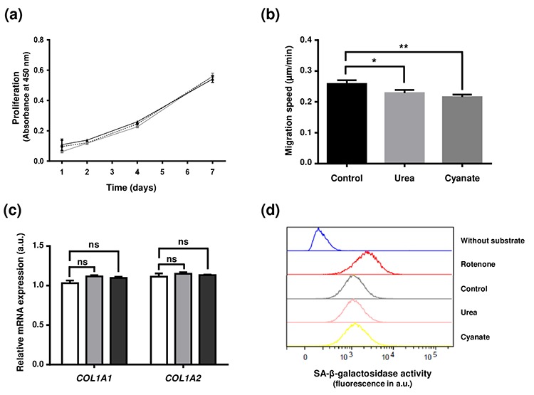
Carbamylation, which corresponds to the binding of isocyanic acid to the amino groups of proteins, is a nonenzymatic post-translational modification responsible for alterations of protein structural and functional properties. Tissue accumulation of carbamylation-derived products and their role in pathological processes such as atherosclerosis or chronic renal failure have been previously documented. However, few studies have focused on the carbamylation of intracellular proteins and their subsequent role in cellular aging. This study aimed to determine the extent of intracellular protein carbamylation, its impact on cell functions and the ability of cells to degrade these modified proteins. Fibroblasts were incubated with cyanate or urea and the carbamylation level was evaluated by immunostaining and homocitrulline quantification. The results showed that carbamylated proteins accumulated intracellularly and that all proteins were susceptible. The presence of intracellular carbamylated proteins did not modify cell proliferation or type I collagen synthesis nor did it induce cell senescence, but it significantly decreased cell motility. Fibroblasts were able to degrade carbamylated proteins through the ubiquitin-proteasome system. In conclusion, intracellular proteins are susceptible to carbamylation but their accumulation does not seem to deeply affect cell function, owing largely to their elimination by the ubiquitin-proteasome system.

Keywords: carbamylation; cell aging; homocitrulline; nonenzymatic post-translational modifications; proteasome; proteostasis.


Conflict of interest statement

CONFLICTS OF INTEREST: The authors do not have any conflict of interest to declare.

Figures

Figure 1
Figure 1
Evidence for the presence of carbamylated proteins inside human skin cells. An anti-HCit immunolabelling was realized in a skin section from a 77-year old man counter-stained with DAPI and hematoxylin-eosin. Epidermal area was screened by light microscopy thanks to hematoxylin-eosin counterstaining (a). Nuclei were highlighted by DAPI staining (b, blue) permitting confirmation of the presence of HCit inside skin cells by immunofluorescence analysis (c, green). An example of a cell exhibiting a positive intracellular labelling for HCit is indicated by arrows. Observations were realized at 10x magnification.
Figure 2
Figure 2
Intracellular accumulation of carbamylated proteins after long-term incubations with urea or cyanate. Confluent fibroblasts were incubated for 4 weeks at 37°C with DMEM + 0.5% (v/v) FBS without (control conditions, open bars) or with 20 mmol/L urea (grey bars) or 0.5 mmol/L cyanate (black bars). HCit content was determined by LC-MS/MS in total cell extracts (a) and in cytoplasmic and membrane fractions (b). The data are presented as means ± SEM (n=6) compared using the Mann-Whitney U test (ns: non significant, **: p<0.01).
Figure 3
Figure 3
Localization of intracellular carbamylated proteins. Fibroblasts were seeded in chambered coverglass system and incubated for 5 days with DMEM containing 0.5% (v/v) FBS and 5 mmol/L cyanate. At the end of incubation, cells were fixed with 4% (v/v) paraformaldehyde and permeabilized with 0.25% (v/v) Triton X-100 before immunolabelling of carbamylated proteins using an anti-HCit polyclonal antibody (a). Cells were also labelled using ActinRed 555 ReadyProbes® in order to identify actin fibers (b). Colocalization points between HCit and actin labelling were identified using ImageJ software (c). In a second set of experiments, fibroblasts were incubated in the same conditions without (control) or with 5 mmol/L cyanate before preparing total cell extracts which were then used for β-actin immunoprecipitation. The immunoprecipitates were submitted to acid hydrolysis before HCit quantification by LC-MS/MS (d). The data are presented as means ± SEM (n=4) compared using the Mann-Whitney U test (**: p<0.01).
Figure 4
Figure 4
Effect of intracellular protein carbamylation on cell function and senescence. Confluent fibroblasts were incubated for 4 weeks at 37°C with DMEM + 0.5% (v/v) FBS without (control conditions) or with 20 mmol/L urea or 0.5 mmol/L cyanate. (a) Proliferation: cells were then seeded in 96-well plates at a density of 1,500 cells per well and incubated for 1, 2, 4 and 7 days with DMEM with 10% (v/v) FBS and carbamylating agents. Cell number was evaluated using a WST-1 assay by measuring absorbance at 450 nm. The data presented are means ± SEM (n=6) compared using the Mann-Whitney U test. No significant difference was found between the three conditions (control: dotted line,•; urea: grey line ■; cyanate: black line, ■). (b) Cell migration: cells were seeded in 24-well plates at a density of 15,000 cells per well and incubated for 24h at 37°C with DMEM containing 0.5% (v/v) FBS. Pictures of cells were taken every 30 min over the incubation period and each cell (n=58) was followed separately in order to calculate the migration speed. The data are presented as means ± SEM compared using the Mann-Whitney U test (*:p<0.05, **:p<0.01). (c) Expression of type I collagen mRNAs: at the end of the 4 weeks-incubation, RNA was isolated from confluent cells and then submitted to RT-qPCR analysis for evaluating the expression of COL1A1 and COL1A2 genes. Data represent the relative mRNA expression normalized to EEF1A1 gene and are expressed as means ± SEM (n=4). The Mann-Whitney U test was used to compare the three conditions: control (open bars), urea (grey bars) and cyanate (black bars). ns: not significant. (d) Senescence: cell senescence was determined by measuring the SA-β-galactosidase activity using a C12FDG fluorogenic substrate and by detection of senescent cells by flow cytometry. Each plot represents the results of 20,000 events acquired per condition. Incubation of cells with rotenone was used as a positive control of cell senescence whereas a negative control without addition of the fluorogenic substrate was performed.
Figure 5
Figure 5
Intracellular degradation of carbamylated proteins. Confluent fibroblasts were incubated for 4 weeks at 37°C with DMEM + 0.5% (v/v) FBS and 0.5 mmol/L cyanate in order to induce intracellular protein carbamylation. Cells were then incubated in the same conditions (with 0.5 mM cyanate, solid line) or without cyanate (dotted line) for two additional weeks. HCit content was determined at each time point. The data are presented as means ± SEM (n=6) and the two conditions (with or without cyanate) were compared using the Mann-Whitney U test (**: p<0.01).
Figure 6
Figure 6
Impact of carbamylation on proteasome proteolytic activities and on the ubiquitination process. (a) Evaluation of proteasome proteolytic activity after incubation of cells with urea or cyanate: confluent fibroblasts were incubated for 4 weeks at 37°C with DMEM + 0.5% (v/v) FBS without (control conditions, open bars) or with 20 mmol/L urea (grey bars) or 0.5 mmol/L cyanate (black bars). Chymotrypsin-like, caspase-like and trypsin-like activities have been measured in cell extracts using the corresponding Proteasome-Glo™ assays. The data are presented as means ± SEM (n=6) and compared using the Mann-Whitney U test (ns: non significant, **: p<0.01). (b) Ubiquitination level of intracellular proteins after incubation of cells with urea or cyanate: confluent fibroblasts were incubated for 4 weeks at 37°C with DMEM + 0.5% (v/v) FBS without (control conditions) or with 20 mmol/L urea or 0.5 mmol/L cyanate, and cell extracts were prepared and submitted to western-blot analysis using an anti-ubiquitin antibody. (c) Anti-HCit and anti-ubiquitin immunolabellings were performed using fibroblasts previously seeded (10,000 cells/well) in chambered coverglass system and incubated for 2 days with DMEM containing 0.5% (v/v) FBS and 5 mmol/L cyanate. At the end of incubation, cells were fixed with 4% (v/v) paraformaldehyde and permeabilized with 0.25% (v/v) Triton X-100 before immunolabelling of proteins using both anti-HCit and anti-ubiquitin antibodies. Colocalization points between HCit and ubiquitin labelling were identified using ImageJ software.
Figure 7
Figure 7
Role of proteasome in the degradation of carbamylated proteins. Confluent fibroblasts were incubated for 7 and 14 days at 37°C with DMEM containing 0.5% (v/v) FBS, 0.5 mmol/L sodium cyanate with or without proteasome inhibitors (10 nmol/L Bortezomib (Bz) or 500 nM MG-132). HCit content in total cell extracts was determined by LC-MS/MS. The data are presented as means ± SEM (n=6) and compared using the Mann-Whitney U test (ns: non significant, **: p<0.01).

References

    1. Kaushik S, Cuervo AM. Proteostasis and aging. Nat Med. 2015; 21:1406–15. 10.1038/nm.4001 - DOI - PubMed
    1. Morimoto RI, Cuervo AM. Proteostasis and the aging proteome in health and disease. J Gerontol A Biol Sci Med Sci. 2014. (Suppl 1); 69:S33–38. 10.1093/gerona/glu049 - DOI - PMC - PubMed
    1. Hipkiss AR. Accumulation of altered proteins and ageing: causes and effects. Exp Gerontol. 2006; 41:464–73. 10.1016/j.exger.2006.03.004 - DOI - PubMed
    1. Golubev A, Hanson AD, Gladyshev VN. Non-enzymatic molecular damage as a prototypic driver of aging. J Biol Chem. 2017; 292:6029–38. 10.1074/jbc.R116.751164 - DOI - PMC - PubMed
    1. Chondrogianni N, Petropoulos I, Grimm S, Georgila K, Catalgol B, Friguet B, Grune T, Gonos ES. Protein damage, repair and proteolysis. Mol Aspects Med. 2014; 35:1–71. 10.1016/j.mam.2012.09.001 - DOI - PubMed

Publication types

MeSH terms

LinkOut - more resources