. 2019 Jun 6;15(6):e1008177.
doi: 10.1371/journal.pgen.1008177. eCollection 2019 Jun.

A mutation in the endonuclease domain of mouse MLH3 reveals novel roles for MutLγ during crossover formation in meiotic prophase I

Affiliations

A mutation in the endonuclease domain of mouse MLH3 reveals novel roles for MutLγ during crossover formation in meiotic prophase I

Melissa Toledo et al. PLoS Genet. .

Abstract

During meiotic prophase I, double-strand breaks (DSBs) initiate homologous recombination leading to non-crossovers (NCOs) and crossovers (COs). In mouse, 10% of DSBs are designated to become COs, primarily through a pathway dependent on the MLH1-MLH3 heterodimer (MutLγ). Mlh3 contains an endonuclease domain that is critical for resolving COs in yeast. We generated a mouse (Mlh3DN/DN) harboring a mutation within this conserved domain that is predicted to generate a protein that is catalytically inert. Mlh3DN/DN males, like fully null Mlh3-/- males, have no spermatozoa and are infertile, yet spermatocytes have grossly normal DSBs and synapsis events in early prophase I. Unlike Mlh3-/- males, mutation of the endonuclease domain within MLH3 permits normal loading and frequency of MutLγ in pachynema. However, key DSB repair factors (RAD51) and mediators of CO pathway choice (BLM helicase) persist into pachynema in Mlh3DN/DN males, indicating a temporal delay in repair events and revealing a mechanism by which alternative DSB repair pathways may be selected. While Mlh3DN/DN spermatocytes retain only 22% of wildtype chiasmata counts, this frequency is greater than observed in Mlh3-/- males (10%), suggesting that the allele may permit partial endonuclease activity, or that other pathways can generate COs from these MutLγ-defined repair intermediates in Mlh3DN/DN males. Double mutant mice homozygous for the Mlh3DN/DN and Mus81-/- mutations show losses in chiasmata close to those observed in Mlh3-/- males, indicating that the MUS81-EME1-regulated crossover pathway can only partially account for the increased residual chiasmata in Mlh3DN/DN spermatocytes. Our data demonstrate that mouse spermatocytes bearing the MLH1-MLH3DN/DN complex display the proper loading of factors essential for CO resolution (MutSγ, CDK2, HEI10, MutLγ). Despite these functions, mice bearing the Mlh3DN/DN allele show defects in the repair of meiotic recombination intermediates and a loss of most chiasmata.


Conflict of interest statement

The authors have declared that no competing interests exist.

Figures
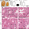
Fig 1
Fig 1. Mlh3DN/DN males show a sterile phenotype.
(A, B) Mlh3DN/DN adult male testes are significantly smaller when compared to WT littermates (Mlh3DN/DN—0.28% of total body weight ± 0.06 standard deviation (s.d.), n = 13; WT—0.77% ± 0.1, n = 15; p < 0.0001, unpaired t-test with Welch's correction) and (C) have zero sperm in the epididymis (WT—9.8 x 10⁷ ± 4.4 sperm/mouse; p < 0.0001; unpaired t-test with Welch's correction, n = 10 and 12 mice, respectively; error bars show standard deviation). Hemotoxylin and eosin stained (D, E) WT testes show the presence of meiotic and post-meiotic cells whereas (F, G) Mlh3DN/DN are absent of spermatids and spermatozoa. Higher magnification of WT and Mlh3DN/DN testes sections are shown in E and G, respectively. Black arrows in (G) indicate metaphase I spermatocytes in the Mlh3DN/DN seminiferous tubule lumen. Sg, spermatogonia; Sc, prophase I spermatocytes; St, postmeiotic spermatids. Scale bar is 100 μm. Images were taken at 40X magnification.
Fig 2
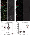
Fig 2. Mlh3DN/DN spermatocytes show a persistence of RAD51 foci in pachynema.
The localization of RAD51 (green) on synaptonemal complex protein SYCP3 (red) is observed in (A, B) WT and (C, D) Mlh3DN/DN male spermatocytes in zygonema and pachynema. (A, C, E, G) In early zygonema, WT cells show high numbers of RAD51 associated with the chromosome cores while Mlh3DN/DN exhibit even higher RAD51 foci (WT mean ± standard deviation = 142.1 ± 34.5 foci, Mlh3DN/DN mean ± s.d. = 188.8 ± 53.3 foci; p values indicated on graph, by unpaired t-test with Welch’s correction, and using Bonferroni’s adjustment for multiple comparison). In late zygonema, Mlh3DN/DN spermatocytes continue to have higher RAD51 foci than WT (WT mean ± s.d. = 193.0 ± 53.7 foci, Mlh3DN/DN mean ± s.d. = 225.3 ± 37.3 foci; p values indicated on graph, by unpaired t-test with Welch’s correction, and using Bonferroni’s adjustment for multiple comparison). Mlh3-/- late zygotene cells show significantly fewer RAD51 focus counts (mean = 122.1 ± 39.9 foci) when compared to WT and Mlh3DN/DN (p values indicated on graph, by unpaired t-test with Welch’s correction, and using Bonferroni’s adjustment for multiple comparison). In all cases, at least 3 mice were assessed for each genotype and at least 20 cells per mouse (B, D, F, H). In pachynema, WT cells exhibit a dramatic decrease of very few to no RAD51 associated with the autosomes while Mlh3DN/DN cells show persistent RAD51 (WT mean ± s.d. = 2.8 ± 2.8 foci, Mlh3DN/DN mean ± s s.d. = 6.1 ± 3.1 foci; p<0.0001, unpaired t-test). Mlh3-/- pachytene cells exhibit comparable RAD51 focus counts (mean ± s.d. = 3.1 ± 2.1 foci) when compared to WT (p = 0.55 by unpaired t-test), but significantly fewer than Mlh3DN/DN spermatocytes (p<0.0001 by unpaired t-test). Note that sex chromosome-associated RAD51 staining was excluded from counts at pachynema. For all chromosome imaging and foci counts, at least three mice of each genotype were observed for each staining set.
Fig 3
Fig 3. Mlh3DN/DN spermatocytes show a persistence of BLM in pachynema.
Meiotic spreads chromosomes from (A-D) WT, (E-H) Mlh3DN/DN, and (I-L) Mlh3-/- males showing the localization of BLM (red) on synaptonemal complex protein SYCP3 (green) throughout the progression of prophase I. (B, F, J, M) In zygonema, Mlh3DN/DN and Mlh3-/- cells show significantly more BLM foci localized to the chromosome cores than WT (WT mean ± s.d. = 221.6 ± 35.1 foci, Mlh3DN/DN mean ± s.d. = 271.1 ± 34.2 foci, Mlh3-/- mean ± s.d. = 265.5 ± 36.6 foci; p values indicated on graph, by unpaired t-test with Welch’s correction, and using Bonferroni’s adjustment for multiple comparison). (C, D, K, M) In pachynema, BLM is no longer present on the chromosome cores of WT cells whereas Mlh3DN/DN and Mlh3-/- cells show hyper-accumulation of BLM on the autosomes and the sex body (WT mean ± s.d. = 7.8 ± 4.7 foci, Mlh3DN/DN mean ± s.d. = 66.7 ± 23.2 foci, Mlh3-/- mean ± s.d. = 58.7 ± 17.8 foci; p values indicated on graph, by unpaired t-test with Welch’s correction, and using Bonferroni’s adjustment for multiple comparison), which persists into diplonema. At zygonema, the frequency of BLM foci in Mlh3DN/DN and Mlh3-/- cells is not statistically different, whereas by pachynema, the number of BLM foci remains significantly elevated in Mlh3DN/DN spermatocytes, relative to that observed in Mlh3-/- cells (p<0.05, unpaired t-test). By diplonema, clear and quantifiable foci are no longer observed in spermatocytes from mice of all genotypes. However, the background staining intensity for these images is observed at standardize exposure settings for image acquisition, so the background staining is comparable between genotypes. For all chromosome imaging and foci counts, at least 3 mice of each genotype were observed for each staining set.
Fig 4
Fig 4. Mlh3DN/DN pachytene spermatocytes exhibit normal localization of MutLγ while the recombinant MLH3-D1185N protein can form complexes with MLH1.
(A-C) MLH3 (green) localizes to SYCP3 (red) in WT and Mlh3DN/DN pachytene spermatocytes with no statistical difference in the number of MLH3 foci (WT = 21.7 ± 3.4 MLH3 foci n = 131, Mlh3DN/DN = 21.3 ± 3.1 MLH3 foci n = 122; p = 0.36 by unpaired t-test). (D-F) MLH1 (green) localizes to SYCP3 (red) in WT and Mlh3DN/DN pachytene spermatocytes also with no statistical difference in the number of MLH1 foci (WT = 21.1 ± 2.6 MLH1 foci n = 144, Mlh3DN/DN = 20.2 ± 2.8 MLH1 foci n = 142; p = 0.2 by unpaired t-test). Different colors in (C) and (F) indicate 3 sets of matched littermates used, each color referring to the counts on a single animal (and color set from the two genotypes being processed simultaneously); n.s = not significant; error bars show standard deviation (s.d.). Note that for MLH1 and MLH3 counts, foci on the pseudoautosomal region (PAR) of the XY bivalent were excluded from quantitation. (G-I) MLH1-mlh3-D1185N forms a stable heterodimer. (G) Schematic of mouse Mlh1 and Mlh3 constructs (see Methods for details). (H) Representative purification of MBP-MLH1-MLH3 (top) and MBP-MLH1-mlh3-D1185N (bottom) using Ni-NTA and amylose resin chromatography as described in the Methods. Fractions were analyzed using SDS-PAGE, stained by Coomassie brilliant blue. MLH1-MLH3 and MLH1-MLH3-D1185N were eluted from amylose in the same fractions. The mass of molecular weight standards is indicated on the left and the expected positions of MBP-MLH1 (127.5 KDa) and His10-MLH3 (165 KDa) is indicated in the center. *Likely to be degradation products of MLH1-MLH3. (I) Mass spectrometry analysis of the two major bands in SDS-PAGE detected after amylose chromatography.
Fig 5
Fig 5. Normal localization of CDK2 and HEI10 to nascent sites of Class I crossovers in Mlh3DN/DN spermatocytes.
(A-D) CDK2 (red) localizes to the synaptonemal complex SYCP3 (green) in WT (A) and Mlh3DN/DN (B) pachytene spermatocytes, but not in Mlh3-/- cells (C), except at the telomeres. Left-hand panels show merged red and green channels, in which white arrows show examples of crossover associated CDK2, while yellow arrows show examples of telomere associated CDK2. Right-hand panels show only the CDK2 signal in white. Panel (D) shows quantitation of foci for each genotype at pachynema. Counts for WT and Mlh3DN/DN are not statistically different from each other (by unpaired t-test with Welch’s correction). Values given are number of foci per nucleus ± s.d. (E-H) HEI10 (red) co-localizes with the synaptonemal complex SYCP3 (green) in pachytene spermatocytes from WT (E), Mlh3DN/DN (F), and Mlh3-/- (G) males. Left-hand panels show merged red and green channels, in which pink arrows show examples of crossover associated HEI10. Right-hand panels show only the HEI10 signal in white. For all chromosome imaging and foci counts, at least 3 mice of each genotype were observed for each staining set. Panel (H) shows quantitation of foci for each genotype at pachynema. Counts for WT and Mlh3DN/DN are not statistically different from each other (by unpaired t-test with Welch’s correction). Values given are number of foci per nucleus ± s.d.
Fig 6
Fig 6
Mlh3DN/DN diakinesis staged spermatocytes show a reduced number of chiasmata (A-D), while comparison of MLH1 focus frequency (E) and distribution (F) during pachynema, and chiasmata counts (G) and bivalent/univalent frequencies (H) in Mus81-/-Mlh3+/+, Mus81-/-Mlh3+/DN, and Mus81-/-Mlh3DN/DN males indicates partial involvement of class II CO pathway. (A-C) Diakinesis staged spermatocyte preparations from WT, Mlh3DN/DN, and Mlh3-/- males stained with Giemsa showing chiasmata formation between homologous chromosomes. Note that diakinesis spread chromosomes are not provided with scale bars because the degree of “spread” varies from cell to cell, thus it is hard to compare true magnifications between each image. Cells were viewed using a 100x objective. (D) Mlh3DN/DN cells exhibit significantly fewer chiasmata when compared to WT (WT = 23.5 ± 1.3 chiasmata per nucleus, Mlh3DN/DN = 5.2 ± 1.7; p values indicated on graph, by unpaired t-test with Welch’s correction, and using Bonferroni’s adjustment for multiple comparisons). Mlh3-/- cells have significantly fewer chiasmata when compared to WT and Mlh3DN/DN (Mlh3-/- = 2.8 ± 1.1 chiasmata per nucleus; p < 0.0001 by unpaired t-test). All values are means ± s.d.. (E-H). We compared MutLγ frequency and distribution during pachynema between mice lacking Mus81 with or without co-incident loss of a endonuclease-intact Mlh3 allele (E, F). Mus81-/-Mlh3+/+ males (E; filled octagons) show elevated MLH1 focus frequency compared to mice bearing one or two copies of the Mlh3DN allele (filled diamonds and down triangles, respectively). The reduced MLH1 focus count in Mus81-/-Mlh3+/DN, and Mus81-/-Mlh3DN/DN males is statistically significant (p values indicated on graph, by unpaired t-test with Welch’s correction, and using Bonferroni’s adjustment for multiple comparisons). (F) The distribution of MLH1 foci is also disrupted in Mus81-/-Mlh3+/DN, and Mus81-/-Mlh3DN/DN males, with increased numbers of synapsed autosomes showing no MLH1 foci, indicative of non-exchange (E0) chromosome pairs, compared to that seen in Mus81-/-Mlh3+/+ males (p values indicated on graph, by unpaired t-test with Welch’s correction, and using Bonferroni’s adjustment for multiple comparison). (G, H) the outcome of class I and class II CO events was assessed by quantifying chiasmata (G) and intact bivalent pairs (H) in diakinesis preparations from mice of all three double mutant genotype combinations, compared to single mutants presented in panel C, above. (G) Chiasmata counts were similar to WT for Mus81-/-Mlh3+/+ and Mus81-/-Mlh3+/DN males (filled octagons and diamonds, respectively), but were statistically significantly lower in Mus81-/-Mlh3DN/DN animals (filled down triangles). The frequency of chiasmata in Mus81-/-Mlh3DN/DN animals was significantly lower than that observed in Mlh3DN/DN animals, but significantly higher than that observed in Mlh3-/- animals (p<0.05, unpaired t-test with Welch’s correction). However, the number of bivalent structures observed at diakinesis in Mus81-/-Mlh3DN/DN animals (panel H) was unchanged from that observed in Mlh3DN/DN animals. In all cases, two animals were assessed for each genotype. Note that for MLH1 and MLH3 counts, foci on the pseudoautosomal region (PAR) of the XY bivalent were excluded from quantitation.

References

    1. Gray S, Cohen PE (2016) Control of meiotic crossovers: from double-strand break formation to designation. Annu Rev Genet 50: 175–210. 10.1146/annurev-genet-120215-035111 - DOI - PMC - PubMed
    1. Hunter N (2015) Meiotic recombination: the essence of heredity. Cold Spring Harb Perspect Biol 7 10.1101/cshperspect.a016618 - DOI - PMC - PubMed
    1. Bolcun-Filas E, Handel MA (2018) Meiosis: the chromosomal foundation of reproduction. Biol Reprod 99: 112–126. 10.1093/biolre/ioy021 - DOI - PubMed
    1. McMahill MS, Sham CW, Bishop DK (2007) Synthesis-dependent strand annealing in meiosis. PLoS Biol 5: e299 10.1371/journal.pbio.0050299 - DOI - PMC - PubMed
    1. Allers T, Lichten M (2001) Intermediates of yeast meiotic recombination contain heteroduplex DNA. Mol Cell 8: 225–231. - PubMed

Publication types