. 2019 Jun 6;14(6):e0218003.
doi: 10.1371/journal.pone.0218003. eCollection 2019.

PINK1 attenuates mtDNA release in alveolar epithelial cells and TLR9 mediated profibrotic responses

Affiliations

PINK1 attenuates mtDNA release in alveolar epithelial cells and TLR9 mediated profibrotic responses

Marta Bueno et al. PLoS One. .

Abstract

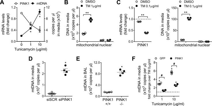
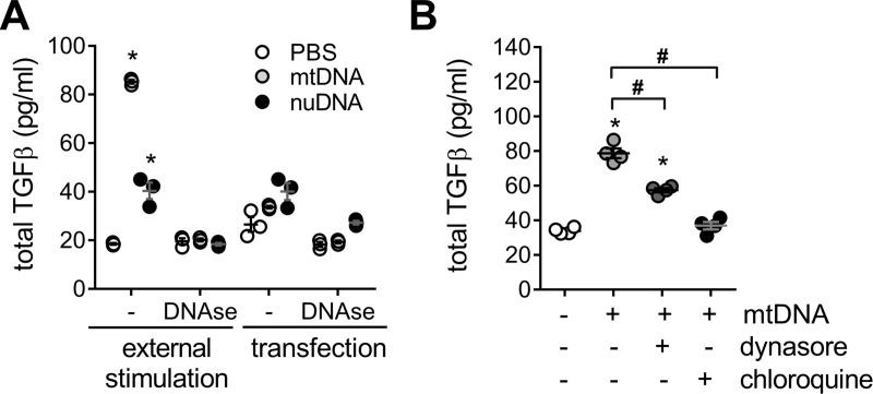
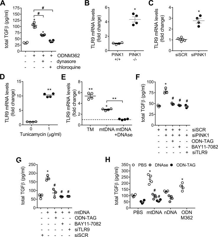
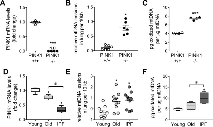
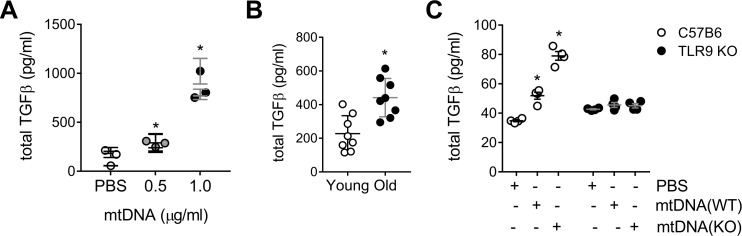
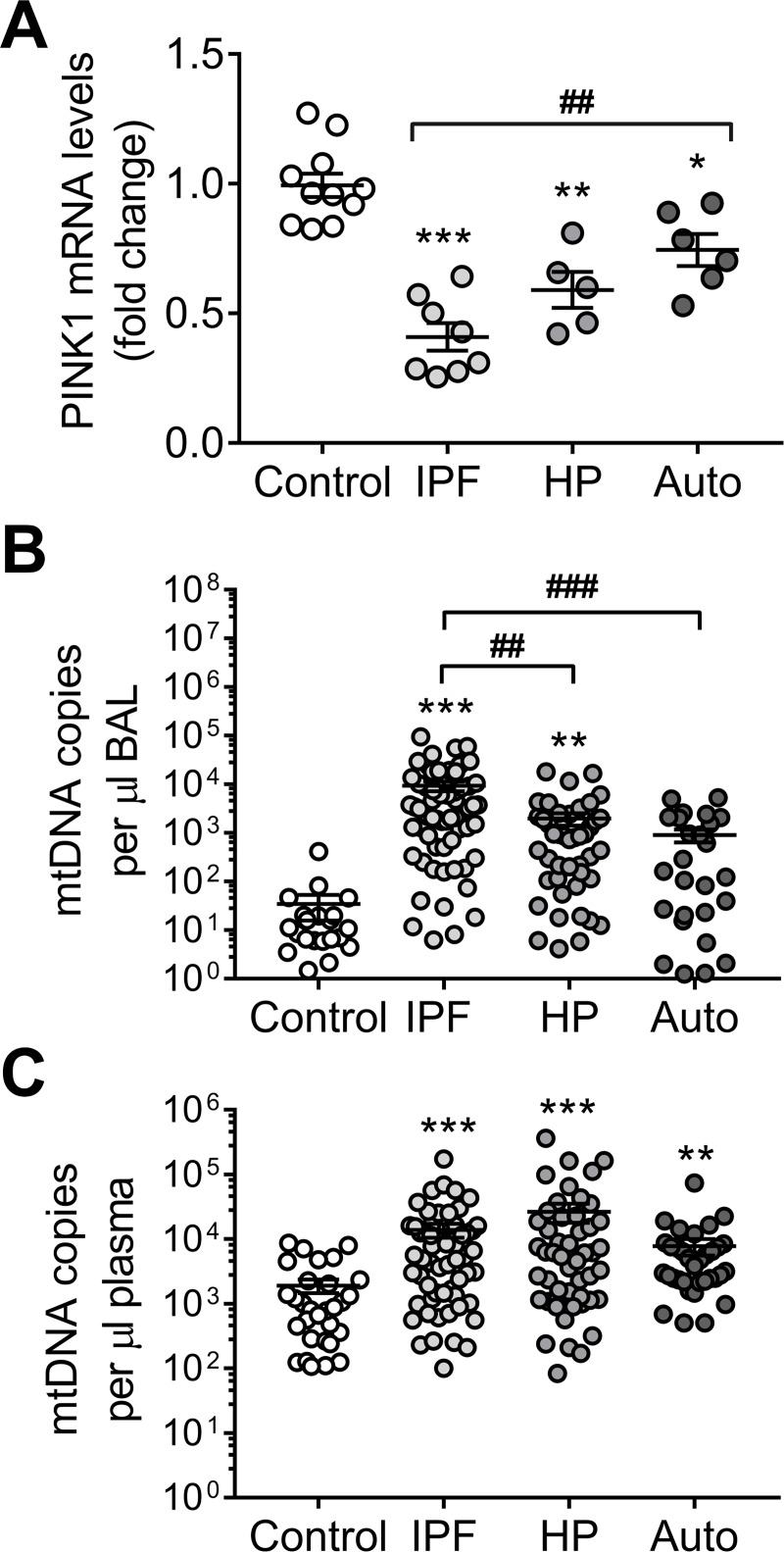
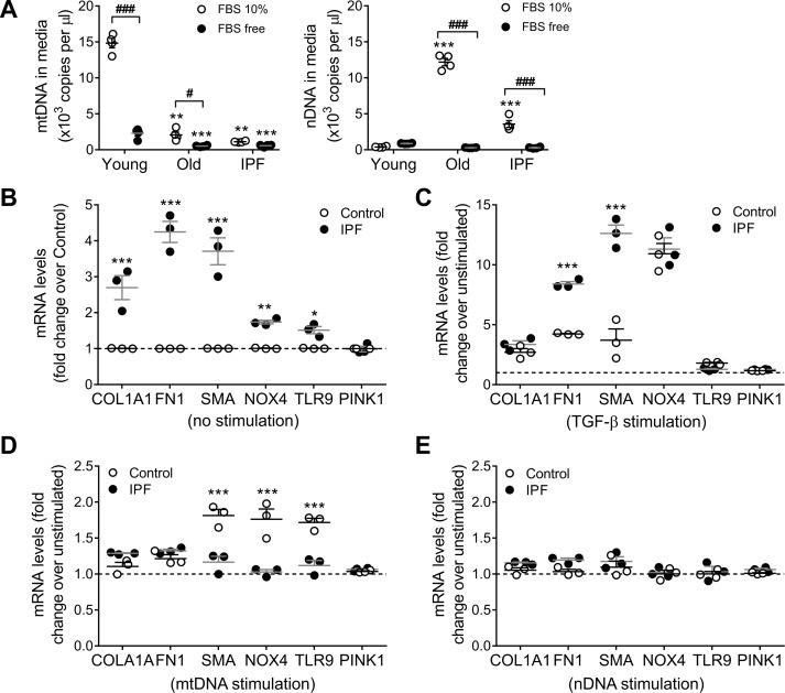
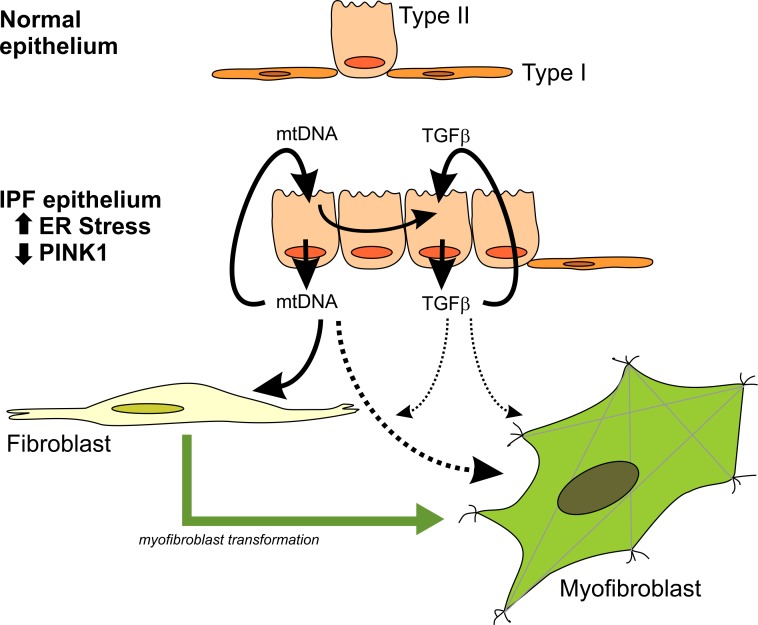
We have previously shown that endoplasmic reticulum stress (ER stress) represses the PTEN inducible kinase 1 (PINK1) in lung type II alveolar epithelial cells (AECII) reducing mitophagy and increasing the susceptibility to lung fibrosis. Although increased circulating mitochondrial DNA (mtDNA) has been reported in chronic lung diseases, the contribution of mitophagy in the modulation of mitochondrial DAMP release and activation of profibrotic responses is unknown. In this study, we show that ER stress and PINK1 deficiency in AECII led to mitochondrial stress with significant oxidation and damage of mtDNA and subsequent extracellular release. Extracellular mtDNA was recognized by TLR9 in AECII by an endocytic-dependent pathway. PINK1 deficiency-dependent mtDNA release promoted activation of TLR9 and triggered secretion of the profibrotic factor TGF-β which was rescued by PINK1 overexpression. Enhanced mtDNA oxidation and damage were found in aging and IPF human lungs and, in concordance, levels of circulating mtDNA were significantly elevated in plasma and bronchoalveolar lavage (BAL) from patients with IPF. Free mtDNA was found elevated in other ILDs with low expression of PINK1 including hypersensitivity pneumonitis and autoimmune interstitial lung diseases. These results support a role for PINK1 mediated mitophagy in the attenuation of mitochondrial damage associated molecular patterns (DAMP) release and control of TGF-β mediated profibrotic responses.


Conflict of interest statement

The authors have declared that no competing interests exist.

Figures

Fig 1
Fig 1. PINK1 deficient lung epithelial cells selectively release mitochondrial DNA.
(A) PINK1 mRNA levels and mtDNA copies in culture media in A549 cells treated with increasing doses of TM for 24h (n = 3; *p<0.01 vs TM 0μg/ml; two-way ANOVA with multiple comparison). (B) Mitochondrial and nuclear DNA copies detected in culture media in A549 cells treated with TM 1μg/ml for 24h (n = 4; **p<0.0001; two-way ANOVA with multiple comparison). (C) PINK1 mRNA levels in primary human alveolar epithelial cells treated 24h with TM 0.1μg/ml (n = 4; **p<0.0001; unpaired t-test). Mitochondrial and nuclear DNA copies detected in culture media in primary human alveolar epithelial cells treated 24h with TM 0.1μg/ml. (n = 4; **p<0.0001; two-way ANOVA with multiple comparison). (D) MtDNA copies in culture media in PINK1 knock-down A549 cells (n = 3, *p<0.001 vs scramble; unpaired t-test). (E) MtDNA copies detected in the BAL fluid of PINK1 +/+ and -/- mice (n = 4, *p<0.001 vs PINK1 +/+; unpaired t-test). (F) MtDNA copies in culture media in A549 cells overexpressing GFP or PINK1 then treated with increasing doses of TM for 24h (n = 4; *p<0.01 vs TM 0μg/ml, #p<0.001 vs GFP; two-way ANOVA with multiple comparison). Dot plots represent mean ± SEM. See S1 Fig for details about ER stress markers levels after TM treatment and knock-down / overexpression efficiencies.
Fig 2
Fig 2. Cells stimulated with free mitochondrial DNA increase TGF-β release.
(A) TGF-β in cell media from A549 treated with extracellular mtDNA or nuclear nDNA (1μg/ml, 24h) and it is absent in the presence of DNAse (1U/ml), using PBS as vehicle (n = 3; *p<0.01 vs PBS; two-way ANOVA with multiple comparison). (B) TFG-β release induced by exogenous mtDNA treatment (1μg/ml, 24h) in A549 cells pretreated for 1h, prior stimulation, with endocytosis blockers (dynasore 80μM or chloroquine 50μM) (n = 4; *p<0.01 vs PBS, #p<0.01; one-way ANOVA with multiple comparison). Dot plots represent mean ± SEM.
Fig 3
Fig 3. TLR9 mediates TGF-β release in response to mitochondrial DNA.
(A) Pre-treatment with endocytosis blockers (dynasore 80μM or chloroquine 50μM) for 1h reduced TFG-β released induced by TRL9 agonist ODN M362 (2μM) after 24h (n = 4; *p<0.01 vs PBS, #p<0.01; one-way ANOVA with multiple comparison). TLR9 mRNA transcript levels in total lung lysate from PINK1 -/- mice (B), A549 cells treated with PINK1 siRNA for 48h (C) or A549 cells treated with 1μg/ml of tunicamycin for 24h (D) (n = 4; *p<0.05, **p<0.01; unpaired t-test). (E) TLR9 mRNA transcript levels in primary human lung epithelial cells (E) after 24 of 0.1μg/ml of tunicamycin or 1 μg/mL exogenous mtDNA in the presence or absence of DNAse (1U/ml) (n = 4; *p<0.05 vs unstimulated, **p<0.01; one-way ANOVA with multiple comparison). TGF-β release in A549 cells were treated with PINK1 siRNA (F) or stimulated with 1 μg/mL exogenous mtDNA for 24h (G) with pre-treatment with a TLR9 antagonist (ODN-TAG, 1μM), the NFκB antagonist BAY11-7082 (5μM), or by silencing TLR9 48h prior stimulation (n = 3; *p<0.001 vs PBS, #p<0.001 vs siPINK1 or mtDNA; one-way ANOVA with multiple comparison). (H) TGF-β release in primary human lung epithelial cells after stimulations with extracellular mtDNA (1μg/ml) in the presence of DNAse (1U/ml), pre-treatments with TRL9 antagonist ODN-TAG (1μM) or exposed to 1μg/ml of nDNA after 24h. TRL9 agonist ODN M362 (1μM) (n = 4; *p<0.001 vs PBS, #p<0.001 vs mtDNA; two-way ANOVA with multiple comparison). Dot plots represent mean ± SEM. See S1C and S1D Fig and S3A Fig for knock-down efficiencies.
Fig 4
Fig 4. IPF and PINK1 deficient lungs show higher mitochondrial DNA oxidation.
(A) PINK1 transcript levels in total lung of PINK1 deficient mice (n = 4; ***p<0.001, N.D non-detected; unpaired t-test). (B) Mitochondrial DNA lesions, by LORD-Q qPCR, in PINK WT and KO mice (n = 6; *p<0.01; unpaired t-test). (C) Oxidative damage in lung mtDNA (by 8-OH-dG per μg of analyzed mtDNA) of PINK1 KO mice (n = 4; ***p<0.001; unpaired t-test). See S4A Fig for aged mice data. (D) Transcript levels of PINK1 in young and old donor lung tissue and in IPF patients. (Min-to-max with median; n = 10; *p<0.01 vs young, #p<0.01; one-way ANOVA with multiple comparison). (E) Mitochondrial DNA lesion by LORD-Q qPCR in lungs of young, old and IPF patients (n = 10; *p<0.01 vs young; one-way ANOVA with multiple comparison). (F) Oxidative damage in lung mtDNA (by 8-OH-dG per μg of analyzed mtDNA) of IPF patients (Min-to-max with median; n = 6; *p<0.01 vs young, #p<0.01; one-way ANOVA with multiple comparison). Dot plots represent mean ± SEM. See S1–S3 Tables for demographics of panel D-F.
Fig 5
Fig 5. Oxidized mtDNA increases downstream production of TGF-β.
(A) TFG-β release in precision-cut lung slices (PCLS) from healthy human donors (60±3) treated with different doses of exogenous mtDNA after 24h (n = 3 patients, n = 6 per patient; *p<0.001 vs PBS; one-way ANOVA with multiple comparison). (B) TFG-β release in human PLCS after 24h of stimulation with 1μg/ml mtDNA isolated from young (35yo) or old (87yo) donor lungs (n = 8 per patient; *p<0.001; unpaired t-test). (C) TGF-β (24h) in C57BL6 or TLR9 mouse PCLS after stimulation with 1μg/ml mtDNA from PINK1+/+ mice (WT) or PINK1 -/- (KO) mice. (n = 4; *p<0.001 vs PBS; two-way ANOVA with multiple comparison). Dot plots represent mean ± SEM. See S5 Fig for representative PCLS viability staining (S5A Fig for human PCLS, S5B Fig for mouse PCLS).
Fig 6
Fig 6. Mitochondrial DNA can be found in the bronchoalveolar lavage (BAL) and the plasma of patients with interstitial lung diseases (ILD).
(A) PINK1 mRNA transcript levels in total lung tissue of aged-matched controls and samples from different ILD (IPF: idiopathic pulmonary fibrosis; HP: hypersensitivity pneumonitis; Auto: autoimmune-related ILD) (n = 10 control, n = 8 IPF, n = 5 HP, n = 6 Auto; *p<0.01, **p<0.001 and ***p<0.0001 vs Control, ##p<0.001; two-way ANOVA with multiple comparisons). See S4 Table for demographic details about this cohort. (B) Detected mtDNA copies by qPCR in the BAL of aged-matched controls and patients with different ILD (n = 22 control, n = 63 IPF, n = 47 HP, n = 29 Auto; **p<0.001 and ***p<0.0001 vs Control, ##p<0.01 and ###p<0.001; two-way ANOVA with multiple comparisons). (C) Detected mtDNA copies by qPCR in the plasma of aged-matched controls and patients with different ILD (n = 30 control, n = 60 IPF, n = 50 HP, n = 35 Auto; **p<0.001 and ***p<0.0001 vs Control; two-way ANOVA with multiple comparisons). Dot plots represent mean ± SEM. See S6B–S6D Fig for details regarding nuclear DNA detection. See S5 Table for demographic details of patients reported in panel B-C.
Fig 7
Fig 7. Lung epithelial cells derived mitochondrial DNA has an effect on lung fibroblast.
(A) Mitochondrial DNA and nuclear release to the media by human lung fibroblast from young (33±4), old age-matched controls (67±3) or IPF patient (68±4) after 24h in culture with different concentrations of FBS (fetal bovine serum) added to the culture media (n = 4; ***p<0.001 and **p<0.01 vs Young, ###p<0.001; two-way ANOVA with multiple comparisons). (B) Baseline expression (non-stimulated) of different pro-fibrotic genes in age-matched human lung fibroblast from control and IPF patients (n = 3; ***p<0.001, **p<0.005, *p< 0.05 vs control; two-way ANOVA with multiple comparisons). (C) Expression of different pro-fibrotic genes in age-matched human lung fibroblast from control and IPF patients after 48h of TGF-β stimulation (5ng/ml) (n = 3; ***p<0.001, vs unstimulated; two-way ANOVA with multiple comparisons). (D) Fold change (over non-stimulated baseline) of pro-fibrotic markers expression after 48h of stimulation with 1μg/ml of exogenous mtDNA in age-matched human lung fibroblast from control and IPF patients (n = 3; ***p<0.001 vs unstimulated; two-way ANOVA with multiple comparisons). (E) Fold change (over non-stimulated baseline) of pro-fibrotic markers expression after 48h of stimulation with 1μg/ml of nuclear DNA in age-matched human lung fibroblast from control and IPF patients (n = 3; two-way ANOVA with multiple comparisons).
Fig 8
Fig 8. Mitochondrial DNA release has a key role in the progression of fibrosis.
Chronically injured, ER-stress sensitive, PINK1-deficient AECII in IPF lungs are the driver to this pro-fibrotic signaling through the release of mtDNA and upregulation of TGFb (via a TLR9-NFκB axis) and it will affect the effector cell (the fibroblast) in different ways. TFGb release could perpetuate the migration of wound healing responding fibroblast to this microenvironment, while mtDNA DAMPs could be key to the progression of the disease by transforming the pool of these fibroblasts to a more pro-fibrotic myofibroblast-like phenotype.

References

    1. Ley B, Collard HR, King TE Jr., Clinical course and prediction of survival in idiopathic pulmonary fibrosis. Am J Respir Crit Care Med. 2011; 183: 431–440. 10.1164/rccm.201006-0894CI - DOI - PubMed
    1. Mora AL, Rojas M, Pardo A, Selman M. Emerging therapies for idiopathic pulmonary fibrosis, a progressive age-related disease. Nat Rev Drug Discov. 2017; 16: 755–772. 10.1038/nrd.2017.170 - DOI - PubMed
    1. Selman M, Pardo A. Revealing the pathogenic and aging-related mechanisms of the enigmatic idiopathic pulmonary fibrosis. an integral model. Am J Respir Crit Care Med. 2014; 189: 1161–1172. 10.1164/rccm.201312-2221PP - DOI - PubMed
    1. Bueno M, Lai YC, Romero Y, Brands J, St Croix CM, Kamga C, et al. PINK1 deficiency impairs mitochondrial homeostasis and promotes lung fibrosis. J Clin Invest. 2015; 125: 521–538. 10.1172/JCI74942 - DOI - PMC - PubMed
    1. Mora AL, Bueno M, Rojas M. Mitochondria in the spotlight of aging and idiopathic pulmonary fibrosis. J Clin Invest. 2017; 127: 405–414. 10.1172/JCI87440 - DOI - PMC - PubMed

Publication types

MeSH terms