Review
. 2019 Sep 14:709:134316.
doi: 10.1016/j.neulet.2019.134316. Epub 2019 Jun 3.

α-Synuclein pathology in Parkinson's disease and related α-synucleinopathies

Affiliations
Review

α-Synuclein pathology in Parkinson's disease and related α-synucleinopathies

Michael X Henderson et al. Neurosci Lett. .

Abstract

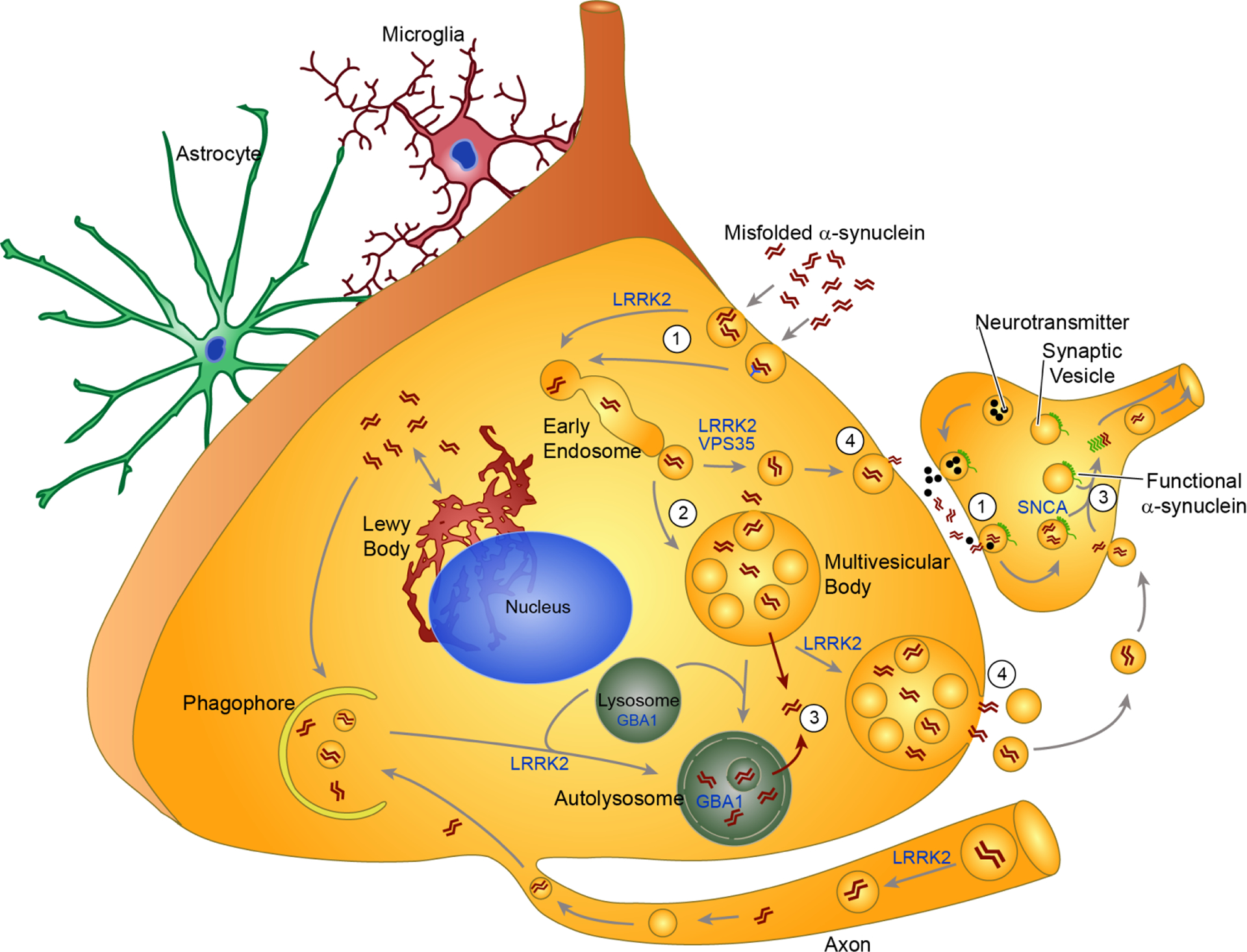
Over 20 years ago, the synaptic protein α-synuclein was identified as the primary component of the Lewy bodies (LBs) that are a sine qua non of Parkinson's disease (PD). Since that time, extensive research has demonstrated that α-synuclein pathology is not only a hallmark of PD, but can also cause neuronal dysfunction and death. Detailed staging of α-synuclein pathology in the brains of patients has revealed a progressive pattern of pathology that correlates with the symptoms of disease. Early in the disease course, PD patients exhibit motor dysfunction, and α-synuclein pathology at this stage is primarily found in regions controlling motor function. At later stages of disease as patients' cognitive function deteriorates, α-synuclein pathology can be found in cortical structures responsible for higher cognitive processing. The stereotypical progression of α-synuclein pathology through the brain over time suggests that there may be a physical transmission of pathological α-synuclein from one area of the brain to another. The transmission hypothesis posits that an initial seed of pathological α-synuclein in one neuron may be released and taken up by another vulnerable neuron and thereby initiate pathological misfolding of α-synuclein in the recipient neuron. In recent years, convergent evidence from various studies has indicated that pathological protein transmission can occur in the human brain. Cell and animal models based on the transmission hypothesis have shown not only that pathological α-synuclein can be transmitted from cell-to-cell, but that this pathology can lead to neuronal dysfunction and degeneration. The α-synuclein transmission hypothesis has profound implications for treatment of what is currently an intractable neurodegenerative disease. In this review, we explore the evidence for cell-to-cell transmission of pathological α-synuclein, the current understanding of how pathological α-synuclein can move to a new cell and template misfolding, and the therapeutic implications of α-synuclein transmission.

Keywords: Lewy body; Misfolding; Neurodegeneration; Prion-like; Transmission.


Figures

FIGURE 1
FIGURE 1
Cell and animal models of alpha-synuclein transmission recapitulate LB aggregates seen in PD. Purified recombinant alpha-synuclein monomer shaken at 37 degrees C and 1000 rotations per minute will spontaneously aggregate into long beta-sheet-rich fibrils. These fibrils can then be sonicated into smaller PFFs that can be added directly to wildtype primary neurons or inoculated into the brain of wildtype mice. This treatment leads to juxtanuclear alpha-synuclein inclusions in neurons and mice that resemble the LBs seen in human PD brains. Cell and mouse inclusions are stained for pS129 alpha-synuclein and the PD tissue is stained for Syn303, which recognizes misfolded alpha-synuclein and reveals LB or LB-like inclusions (blue arrowheads) and Lewy neurites (brown arrowheads). Scale bar = 5 micrometers.
FIGURE 2
FIGURE 2
Mechanisms of pathogenic alpha-synuclein transmission. This schematic of alpha-synuclein transmission demonstrates current hypotheses about the uptake (1), processing (2), escape (3) and release (4) of misfolded alpha-synuclein in a neuron. Astrocytes and microglia are shown in the background because they may also play critical roles in this process. Additionally, several of the shown cellular processes may occur in astrocytes and microglia as well. Extracellular alpha-synuclein can be taken up into neurons either through non-selective endocytosis or receptor-mediated endocytosis (1). In presynaptic terminals, the synaptic vesicle cycle that releases neurotransmitter may also promote uptake of extracellular alpha-synuclein, explaining the retrograde spread of alpha-synuclein pathology in animal models. Following uptake, misfolded alpha-synuclein is processed through the endo-lysosomal pathway (2). At this point, alpha-synuclein may be directly targeted back to the plasma membrane through recycling vesicles, or into multivesicular bodies. At some point in processing, alpha-synuclein seeds can escape the endolysosomal pathway and become free in the cytosol (3). This allows recruitment of endogenous, functional alpha-synuclein into fibrillar structures, a process which would be most devastating at the presynaptic terminal where alpha-synuclein levels are highest (3). Autophagy has the ability to degrade both cytosolic and vesicular alpha-synuclein through engulfment by the phagophore and subsequent fusion with the lysosome. Vesicles or multivesicular bodies containing alpha-synuclein can be targeted for exocytosis, possible to unburden the lysosomal degradation pathway. This leads to release of pathogenic alpha-synuclein from the neuron (4). The LB may provide an additional method of protection for neurons that cannot degrade all misfolded alpha-synuclein. Instead, they deposit it in juxtanuclear inclusions that keep alpha-synuclein from moving around in a more damaging form. Several genetic risk factors for PD encode proteins that are involved in parts of this alpha-synuclein processing pathway. LRRK2 has been implicated in several of the vesicular trafficking pathways. VPS35 may also interact with LRRK2 in vesicle trafficking. GBA1 is a lysosomal enzyme, and mutations may shift the flux through this system.
FIGURE 3
FIGURE 3
Therapeutic implications of alpha-synuclein transmission. The presence of extracellular pathogenic alpha-synuclein species has profound implications for how PD can be treated. This schematic drawing displays several of the therapeutic approaches that are possible to directly address alpha-synuclein transmission. A hypothetical patient is shown with both brain and gut treatments outlined. (1) The recent elucidation of a glymphatic pathway which clears metabolites through connective flow between periarterial and perivenous space presents the possibility of simply enhancing clearance of released alpha-synuclein. This alpha-synuclein would then enter the CSF and potentially meningeal lymph vessels. (2) Immunotherapy targeting alpha-synuclein would exclusively target extracellular species and thereby enhance their degradation through microglia or enhance their clearance into the CSF. (3) Previous work has shown that misfolded alpha-synuclein itself or some factor released by neurons can activate microglia which signal astrocytes and induce a neurotoxic phenotype. Blocking alpha-synuclein binding to microglia, microglial signaling to astrocytes or astrocyte release of neurotoxic factors are all potential therapeutic strategies. (4) Some studies have indicated that extracellular alpha-synuclein is taken up in a receptor-dependent manner and that blockade of binding to the receptor can reduce alpha-synuclein uptake and improve neuron health. (5) PD patients have elevated rates of constipation and deleterious microbiota population. Treatment of constipation via chemical or electrical stimulation or repopulation of intestines with healthy microbiota are treatments which may not only alleviate the GI symptoms experience by PD patients, but also neurological symptoms.

References

    1. Pringsheim T, et al., The prevalence of Parkinson’s disease: a systematic review and meta-analysis. Mov Disord, 2014. 29(13): p. 1583–90. - PubMed
    1. Parkinson J, An essay on the shaking palsy. 1817. J Neuropsychiatry Clin Neurosci, 2002. 14(2): p. 223–36; discussion 222. - PubMed
    1. Poewe W, et al., Parkinson disease. Nat Rev Dis Primers, 2017. 3: p. 17013. - PubMed
    1. Dauer W and Przedborski S, Parkinson’s disease: mechanisms and models. Neuron, 2003. 39(6): p. 889–909. - PubMed
    1. Irwin DJ, Lee VM, and Trojanowski JQ, Parkinson’s disease dementia: convergence of alpha-synuclein, tau and amyloid-beta pathologies. Nat Rev Neurosci, 2013. 14(9): p. 626–36. - PMC - PubMed

Substances