

Development of medical-grade, discrete, multi-walled carbon nanotubes as drug delivery molecules to enhance the treatment of hematological malignancies
- PMID: 31170511
- PMCID: PMC6702103
- DOI: 10.1016/j.nano.2019.102025
Development of medical-grade, discrete, multi-walled carbon nanotubes as drug delivery molecules to enhance the treatment of hematological malignancies
Abstract
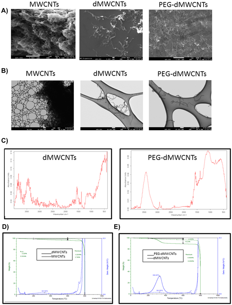
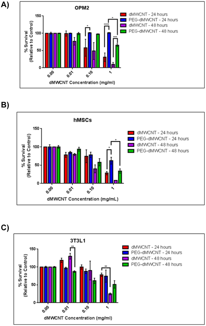
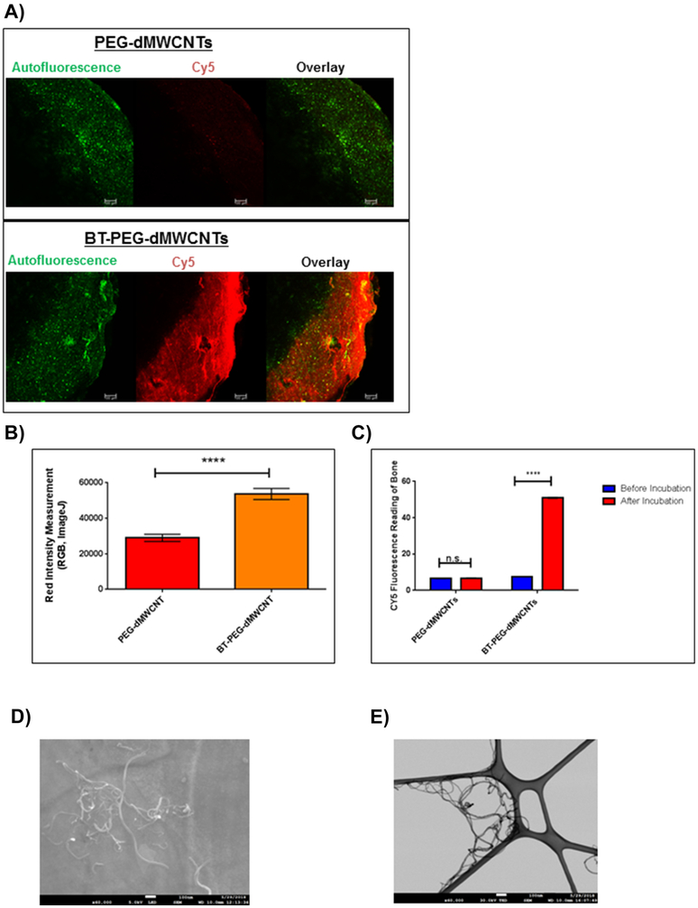
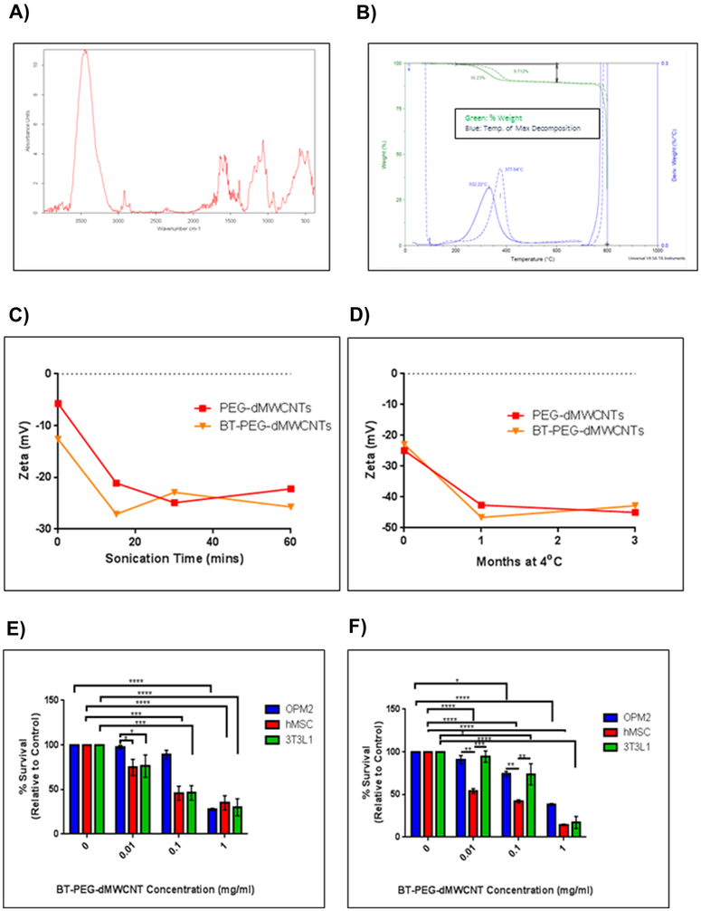
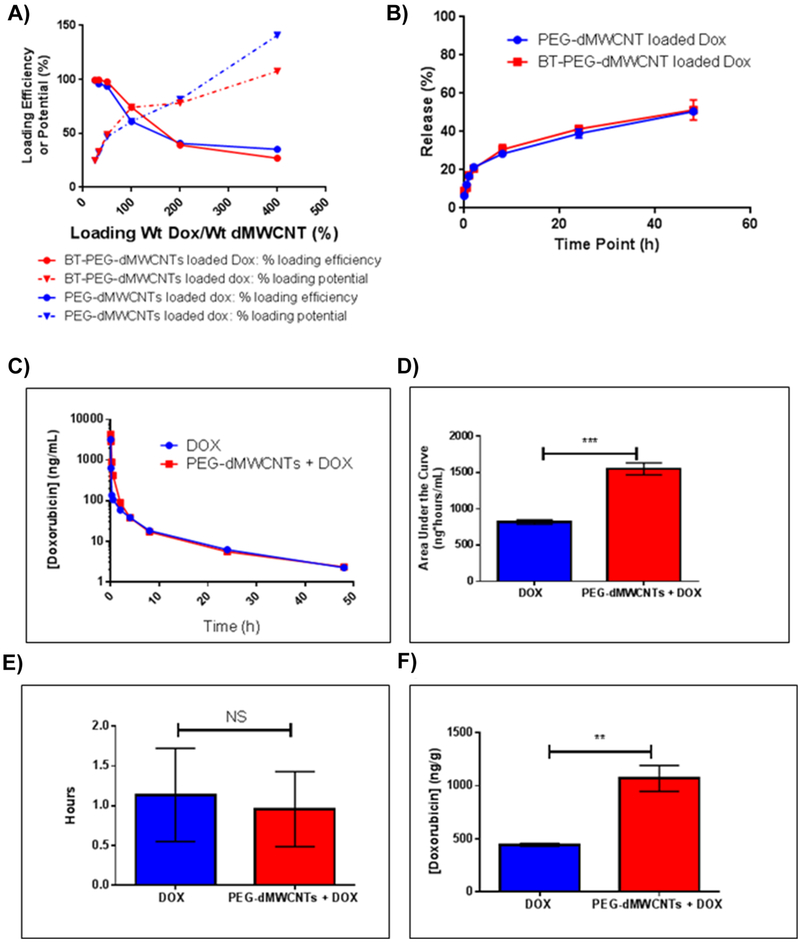
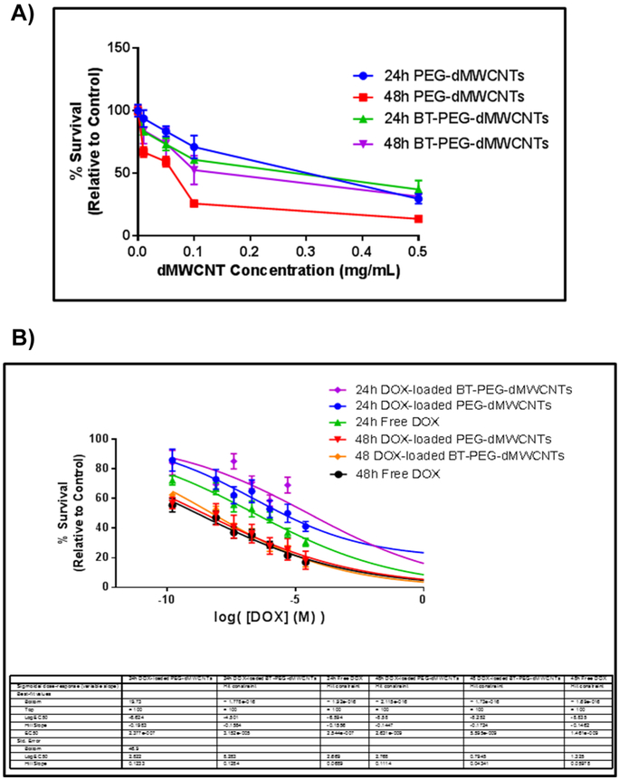
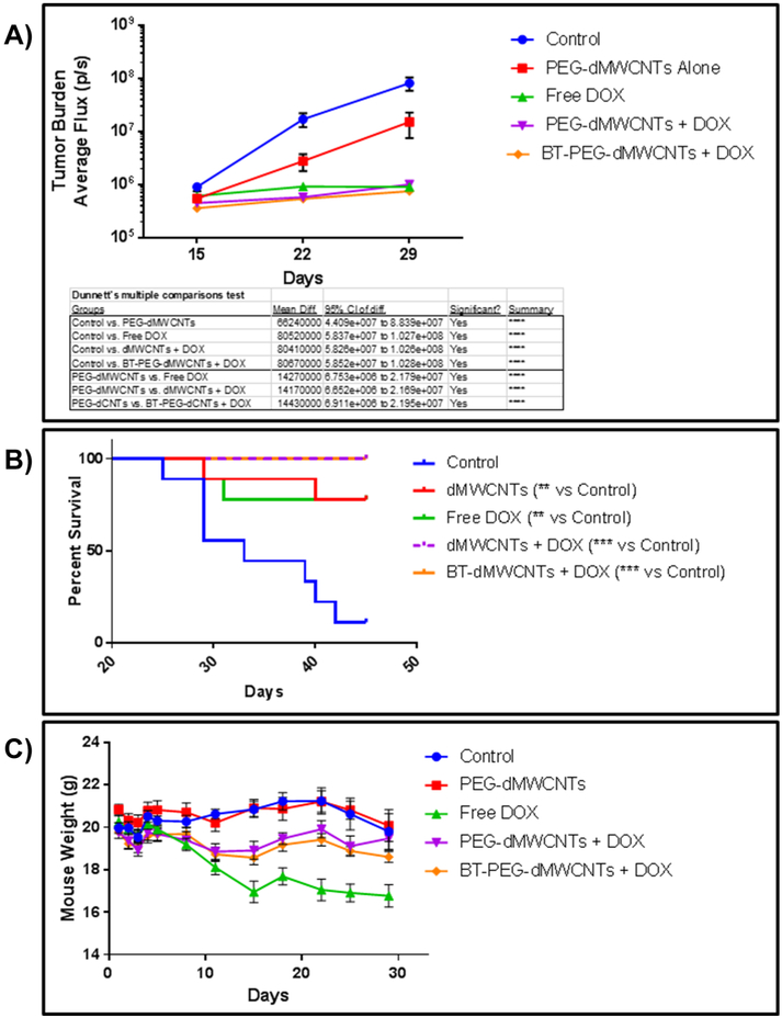
Carbon nanotubes (CNTs) hold great potential as drug delivery transporters given their large drug-binding surface area. Herein, we designed novel, multi-walled, discrete CNTs (dMWCNTs), PEGylated dMWCNTs (PEG-dMWCNTs), and bone-targeting (BT), alendronate-conjugated PEG-dMWCNTs (BT-PEG-dMWCNTs). Using zeta potential, thermogravimetric analysis, TEM, SEM, and FTIR, dMWCNTs were characterized as individual, uniform, and stable. Drug binding and release assays validated dMWCNTs as effective doxorubicin (DOX) transporters. The mass ratio of DOX loading onto dMWCNTs was 35% wt/wt with a ~95% wt/wt efficiency. DOX release was ~51% w/w after 48 hours. Neoplastic transformation, chromosomal aberration, and cytotoxicity assays, confirmed biocompatibility for all dMWCNTs. PEG-dMWCNTs were well tolerated and modulated drug pharmacokinetics in mice. In mice with Burkitt's lymphoma, DOX-loaded PEG-dMWCNTs and BT-PEG-dMWCNTs reduced tumor burden and increased survival similarly to free drug. Importantly, DOX toxicity was abrogated when DOX was loaded onto PEG-dMWCNTs or BT-PEG-dMWCNTs. Overall, PEG-dMWCNTs and BT-PEG-dMWCNTs represent a promising new nanocarrier platform.
Keywords: Burkitt's lymphoma; Discrete carbon nanotubes; Drug delivery; Multiwalled carbon nanotubes; Nanomedicine.
Copyright © 2019 Elsevier Inc. All rights reserved.
Figures
Similar articles
-
PEGylated multi-walled carbon nanotubes as versatile vector for tumor-specific intracellular triggered release with enhanced anti-cancer efficiency: Optimization of length and PEGylation degree.Colloids Surf B Biointerfaces. 2018 Aug 1;168:43-49. doi: 10.1016/j.colsurfb.2018.02.041. Epub 2018 Feb 20. Colloids Surf B Biointerfaces. 2018. PMID: 29482875
-
Folate-conjugated PEG on single walled carbon nanotubes for targeting delivery of Doxorubicin to cancer cells.Macromol Biosci. 2013 Jun;13(6):735-44. doi: 10.1002/mabi.201200475. Epub 2013 Apr 24. Macromol Biosci. 2013. PMID: 23616476
-
The targeted delivery of anticancer drugs to brain glioma by PEGylated oxidized multi-walled carbon nanotubes modified with angiopep-2.Biomaterials. 2012 Apr;33(11):3324-33. doi: 10.1016/j.biomaterials.2012.01.025. Epub 2012 Jan 26. Biomaterials. 2012. PMID: 22281423
-
Carbon nanotubes for delivery of small molecule drugs.Adv Drug Deliv Rev. 2013 Dec;65(15):1964-2015. doi: 10.1016/j.addr.2013.08.005. Epub 2013 Aug 14. Adv Drug Deliv Rev. 2013. PMID: 23954402 Review.
-
PEG-modified carbon nanotubes in biomedicine: current status and challenges ahead.Biomacromolecules. 2011 Oct 10;12(10):3381-93. doi: 10.1021/bm201020h. Epub 2011 Sep 21. Biomacromolecules. 2011. PMID: 21916410 Review.
Cited by
-
Carbon Nanomaterials: Emerging Roles in Immuno-Oncology.Int J Mol Sci. 2023 Apr 1;24(7):6600. doi: 10.3390/ijms24076600. Int J Mol Sci. 2023. PMID: 37047572 Free PMC article. Review.
-
Recent advances in carbon nanomaterials for biomedical applications: A review.Curr Opin Biomed Eng. 2021 Mar;17:100262. doi: 10.1016/j.cobme.2021.100262. Epub 2021 Jan 15. Curr Opin Biomed Eng. 2021. PMID: 33786405 Free PMC article. Review.
-
Carbon Nanomaterials (CNMs) in Cancer Therapy: A Database of CNM-Based Nanocarrier Systems.Pharmaceutics. 2023 May 19;15(5):1545. doi: 10.3390/pharmaceutics15051545. Pharmaceutics. 2023. PMID: 37242787 Free PMC article. Review.
-
Curcumin coating: a novel solution to mitigate inherent carbon nanotube toxicity.J Mater Sci Mater Med. 2024 Mar 25;35(1):24. doi: 10.1007/s10856-024-06789-9. J Mater Sci Mater Med. 2024. PMID: 38526738 Free PMC article.
-
Preparation, characterisation and biological evaluation of biopolymer-coated multi-walled carbon nanotubes for sustained-delivery of silibinin.Sci Rep. 2020 Oct 9;10(1):16941. doi: 10.1038/s41598-020-73963-8. Sci Rep. 2020. PMID: 33037287 Free PMC article.
References
-
- Iijima S. Helical microtubules of graphitic carbon. Lett. To Nat 353, 737–740 (1991).
-
- Teradal NL, Jelinek R. Carbon Nanomaterials in Biological Studies and Biomedicine. Adv. Healthc. Mater , 1700574 (2017). - PubMed
-
- Lalwani G, Patel SC, Sitharaman B. Two- and Three-Dimensional All-Carbon Nanomaterial Assemblies for Tissue Engineering and Regenerative Medicine. Ann. Biomed. Eng 44(6), 1–16 (2016). - PubMed
-
- Arun S, Kanagaraj S. Mechanical characterization and validation of poly (methyl methacrylate)/multi walled carbon nanotube composite for the polycentric knee joint. J. Mech. Behav. Biomed. Mater 50(October 2015), 33–42 (2015). - PubMed
-
- Re F, Moresco R, Masserini M. Nanoparticles for neuroimaging. J. Phys. D. Appl. Phys 45(7), 073001 (2012).
Publication types
MeSH terms
Substances
Grants and funding
LinkOut - more resources
Full Text Sources
