. 2019 Jun 6;19(1):32.
doi: 10.1186/s40644-019-0212-x.

Primary hepatic perivascular epithelioid cell tumors: imaging findings with histopathological correlation

Affiliations

Primary hepatic perivascular epithelioid cell tumors: imaging findings with histopathological correlation

Pei Nie et al. Cancer Imaging. .

Abstract

Background: Hepatic PEComas are very rare. Few systematic reports are available characterizing the imaging and pathological features of hepatic PEComa. The aim of this study was to investigate the imaging findings of primary hepatic perivascular epithelioid cell tumors (PEComa) and its correlation with histopathological features.

Methods: The CT, MRI and ultrasound images and pathological findings of 22 patients with primary hepatic PEComa were retrospectively reviewed.

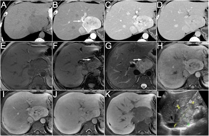
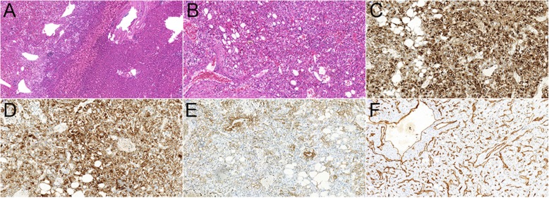
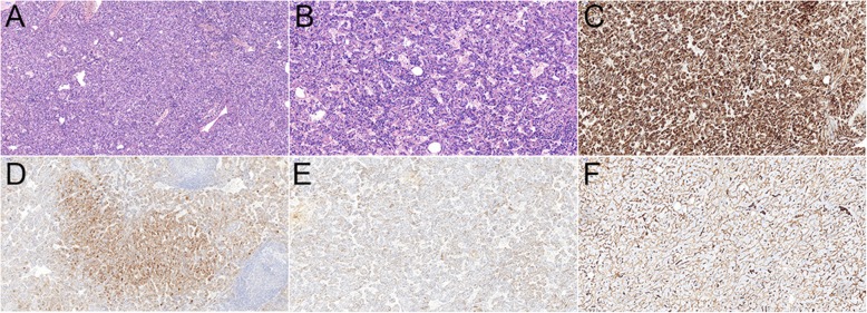
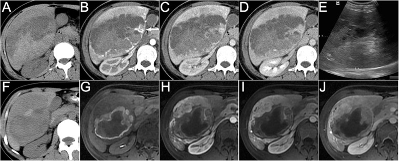
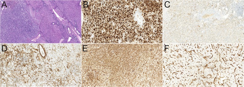
Results: More females (14/22) were affected with the mean age of 47.1 years. Most patients (17/22) were asymptomatic and the routine laboratory tests were normal. More tumors occurred in the right lobe (13/22) with a mean diameter of 76.7 mm. Surgery was performed in 21 patients, and biopsy was performed in 1 patient. Immunohistochemical studies showed the expression rate of HMB-45 and Melan A was 100% (22/22) and 86.4% (19/22) within the tumor cells. The pathology diagnoses were angiomyolipoma (n = 18), lymphangioleiomyoma (n = 2), clear-cell myomelanocytic tumor of falciform ligament/ligamentum teres (n = 1), and not otherwise specified (n = 1). Fifteen cases were classified as uncertain malignant potential (n = 13) or malignant (n = 2). CT, MRI and ultrasound features included well-defined margins (19/22), internal heterogeneity (20/22), arterial enhancement (20/22), dysmorphic vessels (17/22), fat (9/22), hemorrhage (3/22), necrosis (8/22), and calcification (2/22). The diagnostic accuracy was only 27.3% (6/22). No local recurrence or metastasis was found in the follow-up patients (12/22).

Conclusions: On CT, MRI and ultrasound images, most hepatic PEComas are well-defined, heterogeneous, arterial enhanced masses with dysmorphic vessels, with or without fat, especially in middle-aged females. With the potential to be malignant, timely surgical resection and long-term follow-up may be helpful for improving the prognosis.

Keywords: CT; Liver; MRI; Pathology; Perivascular epithelioid cell tumors; Ultrasound.


Conflict of interest statement

The authors declare that they have no competing interest.

Figures

Fig. 1
Fig. 1
A 35-year woman with hepatic angiomyolipoma. The non-enhanced CT (a) showed a round well-circumscribed homogeneous hypoattenuated mass in the caudate hepatic lobe. On the arterial phase (b), the tumor demonstrated significantly heterogeneous enhanced surrounded by a dysmorphic vessel (arrow). On the portal venous (c) and delayed (d) phases, the enhancement decreased, but still showed higher density compared to the surrounding liver. The MRI (e, T1WI in-phase; f, T1WI out-of-phase; g, fat-suppressed T2WI) images showed hypointense on T1WI and hyperintense on T2WI. Note the fat showed signal intensity decreased on out-of-phase image as compared with in-phase image and low signal intensity on fat-suppressed T2WI (arrow). On hepatocyte-specific agent enhanced MRI (h, arterial phase; i, portal venous phase; j, delayed phase; k, hepatobiliary phase), the tumor showed the same enhancement on arterial, portal venous and delayed phase images as CT and marked hypointensity of the tumor relative to the liver parenchyma on the hepatobiliary phase image. Ultrasound (l) showed a well-defined heteroechoic mass in the liver
Fig. 2
Fig. 2
Histological examination of the 35-year woman with hepatic angiomyolipoma. H&E staining images (a, magnification × 100; b magnification × 200) demonstrated that the tumor mainly consisted of three components: the dilated thick-walled vessels; the spindle smooth muscle cells and epitheloid cells in a perivascular location; and some mature lipid tissues. Immunohistochemistry demonstrated the cells were positive for HMB-45 (c, magnification × 200), Melan A (d, magnification × 200), SMA (e, magnification × 200), and CD34 (f, magnification × 200)
Fig. 3
Fig. 3
A 39-year woman with hepatic lymphangioleiomyoma. The non-enhanced CT scan (a) revealed a round well-defined homogeneous hypoattenuated mass in the right hepatic lobe. On the arterial phase (b), the mass demonstrated significantly heterogeneous enhanced surrounded by a dysmorphic vessel (arrow). On the portal venous (c) and delayed (d) phases, the mass became homogeneous, but still showed higher attenuation compared to the background liver. The MRI scan (e, T1WI in-phase; f, T1WI out-of-phase; g, fat-suppressed T2WI; h, DWI) showed hypointense on T1WI, hyperintense on T2WI and high signal intensity on DWI. The enhancement pattern on dynamic contrast-enhanced MRI (i, arterial phase; j, portal venous phase; k, delayed phase) was corresponded with CT. Note the vessel around the tumor on T2WI and arterial phase images (arrow). Ultrasound (l) showed a round, well-defined hyperechoic mass with a cribriform appearance in the right lobe of liver
Fig. 4
Fig. 4
Histological examination of the 39-year woman with hepatic lymphangioleiomyoma. H&E staining images (a, magnification × 100; b magnification × 200) demonstrated that the tumor mainly consisted of the spindle smooth muscle cells and epitheloid cells. Immunohistochemistry demonstrated the cells were positive for HMB-45 (c, magnification × 100), Melan A (d, magnification × 100), SMA (e, magnification × 100) and CD34 (f, magnification × 100)
Fig. 5
Fig. 5
A 29-year woman with clear-cell myomelanocytic tumor of falciform ligament/ligamentum teres. This patient presented acute pain of upper abdomen and was hospitalized in the emergency department. The non-enhanced CT (a) showed a large heterogeneous mass in the right hepatic lobe with high and low attenuation. Contrast-enhanced CT images (b, arterial phase; c, portal venous phase; d, delayed phase) demonstrated heterogeneous enhancement with necrosis and hemorrhage within the mass. Note the mass was surrounded by dysmorphic vessels (arrow) on arterial phase image. Ultrasound (e) showed an ill-defined, large hyperechoic mass with hypoechoic region within the lesion in the right lobe of liver. The patient was performed percutaneous artery embolization. One month later, she underwent CT and contrast-enhanced MRI to reevaluate the mass. Necrosis, hemorrhage and heterogeneous enhancement were also identified on non-enhanced CT (f) and contrast-enhanced MRI (g, mask T1WI; h, arterial phase; i, portal venous phase; j, delayed phase) images. Note the tumor diameter was decreased from 130 mm to 97 mm
Fig. 6
Fig. 6
Histological examination of the 29-year woman with clear-cell myomelanocytic tumor of falciform ligament/ligamentum teres. H&E staining images (a, magnification × 100) demonstrated the dilated thick-walled vessels and perivascular spindled cells and epitheloid cells with clear to lightly eosinophilic cytoplasm. Immunohistochemistry demonstrated the cells were positive for HMB-45 (b, magnification × 200), Melan A (c, magnification × 200), SMA (d, magnification × 200), Vimentin (e, magnification × 200) and CD34 (f, magnification × 200)

References

    1. Son HJ, Kang DW, Kim JH, Han HY, Lee MK. Hepatic perivascular epithelioid cell tumor (PEComa): a case report with a review of literatures. Clin Mol Hepatol. 2017;23:80–86. doi: 10.3350/cmh.2016.0034. - DOI - PMC - PubMed
    1. Tan Y, Zhang H, Xiao EH. Perivascular epithelioid cell tumour: dynamic CT, MRI and clinicopathological characteristics--analysis of 32 cases and review of the literature. Clin Radiol. 2013;68:555–561. doi: 10.1016/j.crad.2012.10.021. - DOI - PubMed
    1. Abhirup B, Kaushal K, Sanket M, Ganesh N. Malignant hepatic perivascular epithelioid cell tumor (PEComa) - case report and a brief review. J Egypt Natl Canc Inst. 2015;27(4):239–242. doi: 10.1016/j.jnci.2015.05.004. - DOI - PubMed
    1. Tirumani SH, Shinagare AB, Hargreaves J, Jagannathan JP, Hornick JL, Wagner AJ, et al. Imaging features of primary and metastatic malignant perivascular epithelioid cell tumors. AJR Am J Roentgenol. 2014;202:252–258. doi: 10.2214/AJR.13.10909. - DOI - PubMed
    1. Ameurtesse H, Chbani L, Bennani A, Toughrai I, Beggui N, Kamaoui I, et al. Primary perivascular epithelioid cell tumor of the liver: new case report and literature review. Diagn Pathol. 2014;9:149. doi: 10.1186/1746-1596-9-149. - DOI - PMC - PubMed

Publication types