Review
. 2019 Sep;27(9):781-791.
doi: 10.1016/j.tim.2019.04.011. Epub 2019 Jun 3.

At the Heart of Bacterial Cytokinesis: The Z Ring

Affiliations
Review

At the Heart of Bacterial Cytokinesis: The Z Ring

Shishen Du et al. Trends Microbiol. 2019 Sep.

Abstract

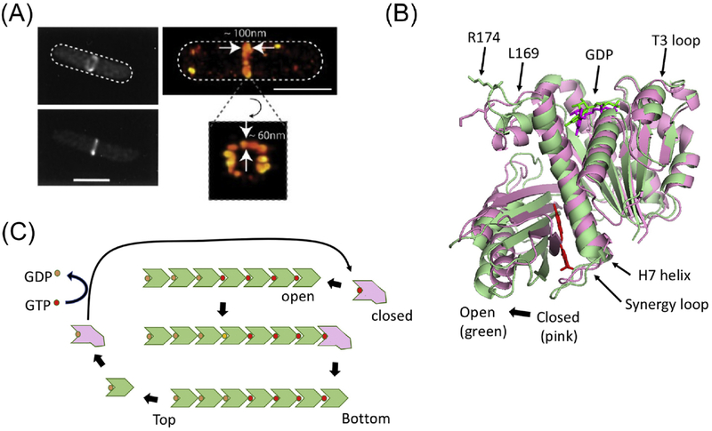
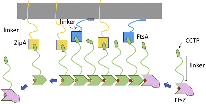
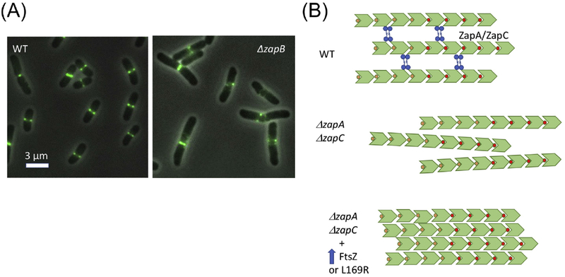
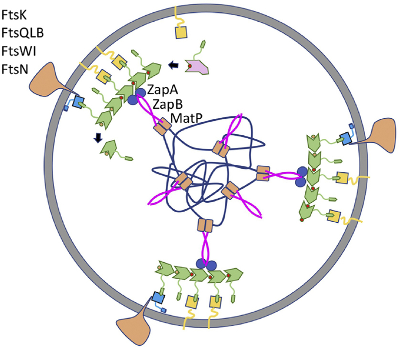
Bacterial cell division is mediated by the divisome which is organized by the Z ring, a cytoskeletal element formed by the polymerization of the tubulin homologue FtsZ. Despite billions of years of bacterial evolution, the Z ring is nearly universal among bacteria that have a cell wall and divide by binary fission. Recent studies have revealed the mechanism of cooperative assembly of FtsZ and that the Z ring consists of patches of FtsZ filaments tethered to the membrane that treadmill to distribute the septal biosynthetic machinery. Here, we summarize these advances and discuss questions raised by these new findings.

Keywords: FtsZ; Z ring; treadmilling; tubulin.


Figures

Figure 1.
Figure 1.. Cooperative assembly of FtsZ.
(A) Conventional and super resolution images of the Z ring (from ref. 14). (B) The two conformations of FtsZ. The open (green – PDB:3VOB) and closed (pink-PDB:2RHL) forms of FtsZ (from Staphylococcus aureus) are superimposed. The PC190723 inhibitor (red) and is present in the structure of the open form. Residues corresponding to R174 and L169 are indicated as are other important features. (C) A model for the cooperative assembly of FtsZ. In this model FtsZ in the closed form adds to the bottom end (the kinetic plus end) of a filament and switches conformation to the open form. Subsequent GTP hydrolysis leads to weakening of longitudinal bonds and loss of subunits from the minus end. Such subunits revert to the closed form and undergo nucleotide exchange before adding to the growing end.
Figure 2.
Figure 2.. Linking FtsZ filaments to the membrane.
FtsZ filaments bind to membrane anchors (FtsA and ZipA). This binding involves the CCTP (conserved C-terminal peptide) of FtsZ binding to FtsA and ZipA. The CCTP is an example of a MoRF (molecular recognition feature) more common in eukaryotic proteins. Such motifs display weak affinity and this is true for the CCTP binding to FtsA or ZipA, however, an FtsZ filament displays strong binding due to avidity (many CCTPs binding many membrane anchors). The intrinsically disordered regions (linker) in FtsZ, ZipA and FtsA increase flexibility allowing interaction with their partners. The long linker in ZipA may help it snare FtsZ filaments while the long linker in FtsZ with the CCTP at the tip may function like a grappling hook.
Figure 3.
Figure 3.. Bundling vs cross-linking of FtsZ filaments.
(A) Images of FtsZ-GFP in wild type (WT) and a mutant lacking the Ter signal (ΔzapB). (B) FtsZ filaments are present in patches in the Z ring. The filaments are presumed to be aligned in parallel based upon the directional movement seen in bundles and patches. In WT cells the primary interaction between filaments may be due to crosslinking proteins (ZapA, ZapC, and ZapD). In the absence of two of these (ZapA and ZapC) cells are elongated and cells contain spiral Z rings (similar to images in (A)). Overexpression of FtsZ or the presence of FtsZ-L169R may lead to increased bundling that compensates for the loss of Zap proteins.
Figure 4.
Figure 4.. Cell wall synthesis distributed by treadmilling FtsZ filaments linked to Ter.
FtsZ filaments attached to the membrane assemble into the Z ring (FtsZ, FtsA and ZipA colored as in Fig. 2), which consists of patches of FtsZ filaments that move circumferentially around the septum in both directions. Assembly at midcell is aided by the presence of the Ter signal consisting of ZapA bound to FtsZ linked to the MatP macrodomain by ZapB. The septal peptidoglycan synthetic machinery (depicted as a single large complex) consists of enzymes (FtsW-transglcosylase and Ftsl-transpeptidase) and proteins involved in their recruitment (FtsK, FtsQLB) and regulation (FtsQLB, FtsN).

References

    1. Nanninga N (1991) Cell division and peptidoglycan assembly in Escherichia coli. Mol Microbiol 5 (4), 791–5. - PubMed
    1. Lutkenhaus J (1993) FtsZ ring in bacterial cytokinesis. Mol Microbiol 9 (3), 403–9. - PubMed
    1. Haeusser DP and Margolin W (2016) Splitsville: structural and functional insights into the dynamic bacterial Z ring. Nat Rev Microbiol 14 (5), 305–19. - PMC - PubMed
    1. Du S and Lutkenhaus J (2017) Assembly and activation of the Escherichia coli divisome. Mol Microbiol 105 (2), 177–187. - PMC - PubMed
    1. Lutkenhaus J et al. (2012) Bacterial cytokinesis: From Z ring to divisome. Cytoskeleton (Hoboken) 69 (10), 778–90. - PMC - PubMed

Publication types

MeSH terms

LinkOut - more resources