

A Fully Automatic Technique for Precise Localization and Quantification of Amyloid-β PET Scans
- PMID: 31171596
- PMCID: PMC6894379
- DOI: 10.2967/jnumed.119.228510
A Fully Automatic Technique for Precise Localization and Quantification of Amyloid-β PET Scans
Abstract
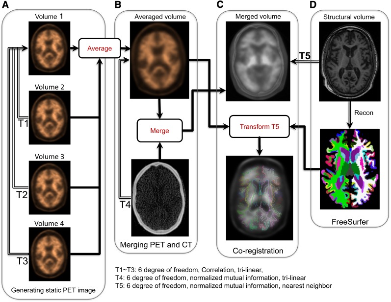
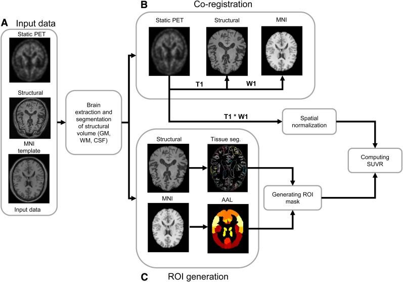
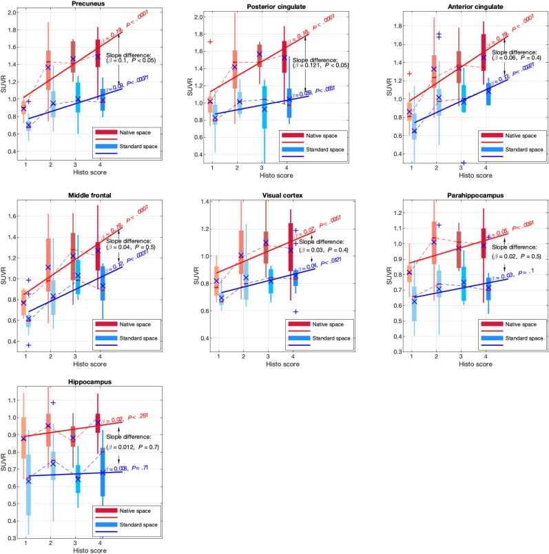
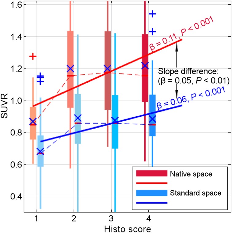
Spatial heterogeneity in the accumulation of amyloid-β plaques throughout the brain during asymptomatic as well as clinical stages of Alzheimer disease calls for precise localization and quantification of this protein using PET imaging. To address this need, we have developed and evaluated a technique that quantifies the extent of amyloid-β pathology on a millimeter-by-millimeter scale in the brain with unprecedented precision using data from PET scans. Methods: An intermodal and intrasubject registration with normalized mutual information as the cost function was used to transform all FreeSurfer neuroanatomic labels into PET image space, which were subsequently used to compute regional SUV ratio (SUVR). We have evaluated our technique using postmortem histopathologic staining data from 52 older participants as the standard-of-truth measurement. Results: Our method resulted in consistently and significantly higher SUVRs in comparison to the conventional method in almost all regions of interest. A 2-way ANOVA revealed a significant main effect of method as well as a significant interaction effect of method on the relationship between computed SUVR and histopathologic staining score. Conclusion: These findings suggest that processing the amyloid-β PET data in subjects' native space can improve the accuracy of the computed SUVRs, as they are more closely associated with the histopathologic staining data than are the results of the conventional approach.
Keywords: 18F-florbetaben PET; Alzheimer disease; FreeSurfer; amyloid-β; spatial normalization.
© 2019 by the Society of Nuclear Medicine and Molecular Imaging.
Figures
References
-
- Seibyl J, Catafau AM, Barthel H, et al. Impact of training method on the robustness of the visual assessment of 18F-florbetaben PET scans: results from a phase-3 study. J Nucl Med. 2016;57:900–906. - PubMed
-
- Joshi AD, Pontecorvo MJ, Lu M, Skovronsky DM, Mintun MA, Devous MDS. A semiautomated method for quantification of F 18 florbetapir PET images. J Nucl Med. 2015;56:1736–1741. - PubMed
-
- Zubal G, Wisniewski G, Seibyl J. Automated software package for analyzing new beta-amyloid radioligands in Alzheimer’s patients [abstract]. J Nucl Med. 2008;49(suppl):378P–378P.
-
- Fleisher AS. Using positron emission tomography and florbetapir F 18 to image cortical amyloid in patients with mild cognitive impairment or dementia due to Alzheimer disease. Arch Neurol. 2011;68:1404–1411. - PubMed
Publication types
MeSH terms
Substances
Grants and funding
LinkOut - more resources
Full Text Sources