. 2019 Nov;60(11):1569-1578.
doi: 10.2967/jnumed.119.225813. Epub 2019 Jun 6.

Temporal Modulation of HER2 Membrane Availability Increases Pertuzumab Uptake and Pretargeted Molecular Imaging of Gastric Tumors

Affiliations

Temporal Modulation of HER2 Membrane Availability Increases Pertuzumab Uptake and Pretargeted Molecular Imaging of Gastric Tumors

Patrícia M R Pereira et al. J Nucl Med. 2019 Nov.

Abstract

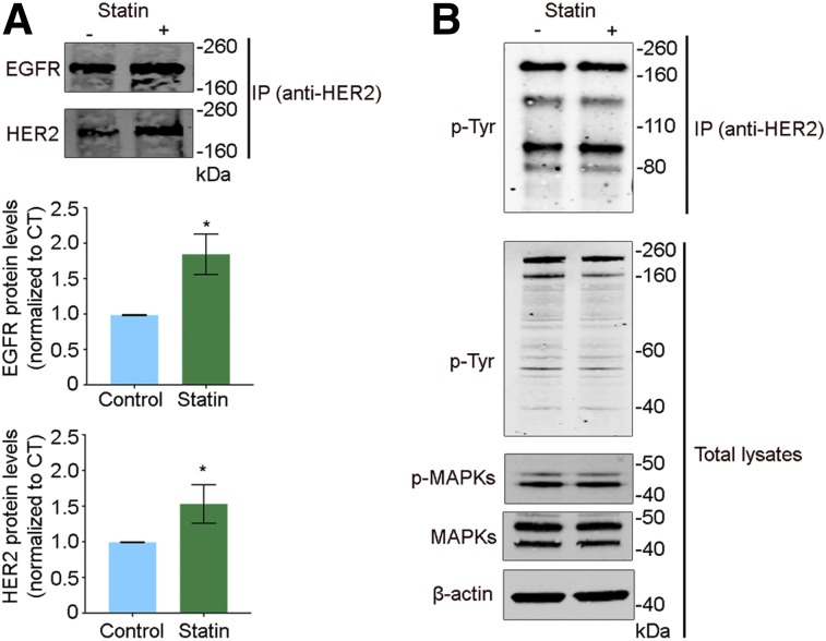
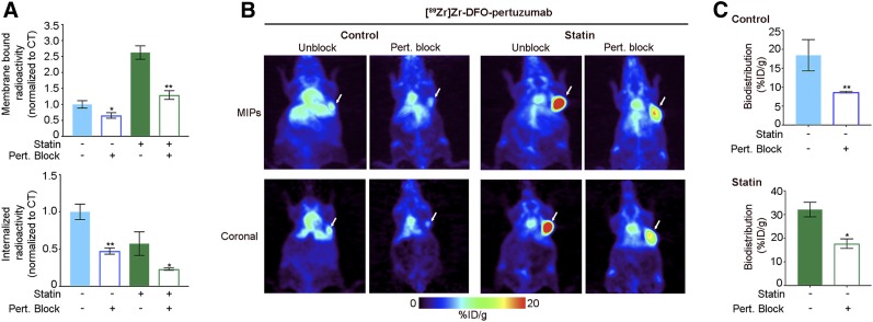
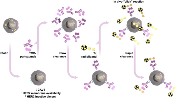
Human epidermal growth factor receptor 2 (HER2) is used as a tumor biomarker and therapeutic target. Pertuzumab is an anti-HER2 antibody, and its binding to tumor cells requires HER2 to be present at the cell membrane. However, the cellular distribution of HER2 protein in gastric tumors is dynamic, and HER2 internalization decreases antibody binding to tumor cells. These features preclude the use of pretargeted strategies for molecular imaging and therapy. We explored the pharmacological modulation of HER2 endocytosis as a strategy to improve pertuzumab uptake in HER2-positive gastric tumors and allow the use of a pretargeted imaging approach. Methods: We conducted in vitro and in vivo studies with NCI-N87 gastric cancer cells to determine how HER2 endocytosis affects pertuzumab binding to tumor cells. Lovastatin, a clinically approved cholesterol-lowering drug, was used to modulate caveolae-mediated HER2 endocytosis. Results: Administration of lovastatin to NCI-N87 cancer cells resulted in significant accumulation of non-activated HER2 dimers at the cell surface. Pretreatment of NCI-N87 cells with lovastatin increased in vitro specific accumulation of membrane-bound 89Zr-labeled pertuzumab. Lovastatin-enhanced pertuzumab tumor uptake was also observed in NCI-N87 gastric cancer xenografts, allowing tumor detection as early as 4 h and high-contrast images at 48 h after tracer administration via PET. Temporal enhancement of HER2 membrane availability by lovastatin allowed imaging of cell surface HER2 with transcyclooctene-conjugated antibodies and 18F-labeled tetrazine. Conclusion: Temporal pharmacological modulation of membrane HER2 may be clinically relevant and exploitable for pretargeted molecular imaging and therapy in gastric tumors.

Keywords: HER2; gastric tumors; lovastatin; pertuzumab; pretargeting.


Figures

FIGURE 1.
FIGURE 1.
Lovastatin treatment increases HER2–HER2 and HER2–EGFR non-activated dimers in gastric cancer cells. Western blots are shown for EGFR, HER2, phosphorylated tyrosine (p-Tyr), phosphorylated MAPKs (p-MAPKs), and total MAPKs from total cell extracts or extracts obtained after immunoprecipitation (IP) with anti-HER2 antibody. Untreated NCI-N87 gastric cancer cells served as control. NCI-N87 cells were incubated with 25 μM lovastatin for 4 h. β-actin was used as loading control. Western blot quantifications of EGFR and HER2 (normalized to control) are represented as mean ± SEM (*P < 0.05 based on Student t test). Experiment was repeated 3 times.
FIGURE 2.
FIGURE 2.
Lovastatin treatment increases in vitro membrane-bound pertuzumab and in vivo tumors’ avidity for pertuzumab. (A) Membrane-bound and internalized [89Zr]Zr-DFO-pertuzumab after treatment with lovastatin in NCI-N87 cancer cells. NCI-N87 untreated cells served as control. NCI-N87 cancer cells were incubated with 25 μM lovastatin for 4 h before addition of 1 μM 89Zr-labeled pertuzumab for 1.5 h. Data represent mean ± SEM (n = 4 experiments, *P < 0.05 based on Student t test). (B and C) Biodistribution (B) and representative maximum-intensity-projection images (MIPs) and coronal PET images (C) of [89Zr]Zr-DFO-pertuzumab in athymic nude mice bearing subcutaneous NCI-N87 gastric tumors treated with lovastatin. Lovastatin (8.3 mg/kg of mouse) was orally administered 12 h before and at same time as tail vein injection of [89Zr]Zr-DFO-pertuzumab (4.44–5.18 MBq, 42–49 μg of protein). Control mice received oral saline instead of lovastatin. Biodistribution data represent mean ± SEM (n = 5 mice per group, *P < 0.05 based on Student t test).
FIGURE 3.
FIGURE 3.
Lovastatin-mediated increase in membrane-bound pertuzumab is blocked with excess of pertuzumab. (A) Membrane-bound and internalized [89Zr]Zr-DFO-pertuzumab in presence of excess of unlabeled pertuzumab after lovastatin treatment in NCI-N87 cells. Blocking experiments were performed by incubation with 89Zr-labeled pertuzumab in presence of 30-fold excess of pertuzumab. Untreated NCI-N87 cells served as control. NCI-N87 cells were incubated with 25 μM lovastatin for 4 h before addition of 1 μM 89Zr-labeled pertuzumab and incubation for 1.5 h. Data represent mean ± SEM (n = 4 experiments, *P < 0.05 and **P < 0.01 based on Student t test). (B and C) Representative maximum-intensity-projection images (MIPs) and coronal PET images (B) and tumor uptake (at 48 h after injection of 89Zr-labeled pertuzumab) (C) in athymic nude mice bearing subcutaneous NCI-N87 gastric tumors with and without blocking with unlabeled pertuzumab. Lovastatin (8.3 mg/kg) was orally administered 12 h before and at same time as tail vein injection of [89Zr]Zr-DFO-pertuzumab (4.44–5.18 MBq, 42–49 μg of protein). Control mice received oral saline. Blocking experiments were performed by administration of 89Zr-labeled pertuzumab in presence of 40-fold excess of pertuzumab. Data represent mean ± SEM (n = 5 mice per group, *P < 0.05 and **P < 0.01 based on Student t test).
FIGURE 4.
FIGURE 4.
Trastuzumab-receptor blockade increases internalization of 89Zr-labeled pertuzumab. (A) Membrane-bound and internalized [89Zr]Zr-DFO-pertuzumab in presence of trastuzumab in NCI-N87 cancer cells with or without lovastatin treatment. Blocking experiments were performed by incubation with 89Zr-labeled pertuzumab in presence of 30-fold excess of trastuzumab. NCI-N87 untreated cells served as control. NCI-N87 cancer cells were incubated with 25 μM lovastatin for 4 h before addition of 1 μM 89Zr-labeled pertuzumab for 1.5 h. Data represent mean ± SEM (n = 4 experiments, *P < 0.05 based on Student t test). (B and C) Representative coronal PET images (B) and tumor uptake (at 48 h after injection of 89Zr-labeled pertuzumab) (C) in athymic nude mice bearing subcutaneous NCI-N87 gastric tumors with and without blocking with unlabeled trastuzumab. Lovastatin (8.3 mg/kg of mice) was orally administered 12 h before and at same time as tail vein injection of [89Zr]Zr-DFO-pertuzumab (4.44–5.18 MBq, 42–49 μg of protein). Control mice received oral saline. Blocking experiments were performed by administration of 89Zr-labeled pertuzumab in presence of 40-fold excess of trastuzumab. Data represent mean ± SEM (n = 5 mice per group, ***P < 0.001 based on Student t test). MIP = maximum-intensity projection.
FIGURE 5.
FIGURE 5.
Schematic of pretargeting approach to image gastric tumors with pertuzumab in presence of lovastatin. Lovastatin depletes CAV1, increasing HER2 membrane availability and HER2 inactive dimers for binding pertuzumab. TCO-labeled pertuzumab (slow pharmacokinetics) is injected days ahead of administering radiolabeled small molecule. Then, only hours before imaging, administered radiolabeled small molecule travels through blood rapidly, either clicking with TCO-labeled antibody or quickly clearing from patient. HER2 is represented in light blue and CAV1 in yellow.
FIGURE 6.
FIGURE 6.
Pretreatment of gastric tumor cells with lovastatin improves pretargeted molecular imaging. Shown are representative coronal PET images (A), maximum-intensity-projection images (MIPs) (B), and biodistribution (C) at 4 h after injection of [18F]AlF-NOTA-PEG11-Tz in athymic nude mice bearing subcutaneous gastric tumors. Lovastatin (8.3 mg/kg of mouse) was orally administered 12 h before and at same time as tail vein injection of pertuzumab-TCO. Mice were administered pertuzumab-TCO (0.42 nmol) 24 h before injection of 18F-labeled tracer (14.73–16.54 MBq, 0.83 nmol) via tail vein. Data represent mean ± SEM (n = 5 experiments).

References

    1. Yarden Y, Sliwkowski MX. Untangling the ErbB signalling network. Nat Rev Mol Cell Biol. 2001;2:127–137. - PubMed
    1. Ocana A, Vera-Badillo F, Seruga B, Templeton A, Pandiella A, Amir E. HER3 overexpression and survival in solid tumors: a meta-analysis. J Natl Cancer Inst. 2013;105:266–273. - PubMed
    1. Hedner C, Borg D, Nodin B, Karnevi E, Jirstrom K, Eberhard J. Expression and prognostic significance of human epidermal growth factor receptors 1, 2 and 3 in oesophageal and gastric adenocarcinomas preneoadjuvant and postneoadjuvant treatment. J Clin Pathol. 2018;71:451–462. - PubMed
    1. Citri A, Yarden Y. EGF-ERBB signalling: towards the systems level. Nat Rev Mol Cell Biol. 2006;7:505–516. - PubMed
    1. Tzahar E, Pinkas-Kramarski R, Moyer JD, et al. Bivalence of EGF-like ligands drives the ErbB signaling network. EMBO J. 1997;16:4938–4950. - PMC - PubMed

Publication types

MeSH terms