Review
. 2019 Jul 19;294(29):11062-11086.
doi: 10.1074/jbc.REV119.005601. Epub 2019 Jun 5.

Illuminating the Onco-GPCRome: Novel G protein-coupled receptor-driven oncocrine networks and targets for cancer immunotherapy

Affiliations
Review

Illuminating the Onco-GPCRome: Novel G protein-coupled receptor-driven oncocrine networks and targets for cancer immunotherapy

Victoria Wu et al. J Biol Chem. .

Abstract

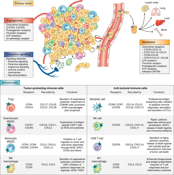
G protein-coupled receptors (GPCRs) are the largest gene family of cell membrane-associated molecules mediating signal transmission, and their involvement in key physiological functions is well-established. The ability of GPCRs to regulate a vast array of fundamental biological processes, such as cardiovascular functions, immune responses, hormone and enzyme release from endocrine and exocrine glands, neurotransmission, and sensory perception (e.g. vision, odor, and taste), is largely due to the diversity of these receptors and the layers of their downstream signaling circuits. Dysregulated expression and aberrant functions of GPCRs have been linked to some of the most prevalent human diseases, which renders GPCRs one of the top targets for pharmaceutical drug development. However, the study of the role of GPCRs in tumor biology has only just begun to make headway. Recent studies have shown that GPCRs can contribute to the many facets of tumorigenesis, including proliferation, survival, angiogenesis, invasion, metastasis, therapy resistance, and immune evasion. Indeed, GPCRs are widely dysregulated in cancer and yet are underexploited in oncology. We present here a comprehensive analysis of GPCR gene expression, copy number variation, and mutational signatures in 33 cancer types. We also highlight the emerging role of GPCRs as part of oncocrine networks promoting tumor growth, dissemination, and immune evasion, and we stress the potential benefits of targeting GPCRs and their signaling circuits in the new era of precision medicine and cancer immunotherapies.

Keywords: G protein; G protein–coupled receptor (GPCR); cancer; drug repurposing; immunotherapy; oncocrine signaling; precision therapies; signal transduction.


Conflict of interest statement

J. S. G. is a member of the Scientific Advisory Board of Oncoceutics Inc. and Domain Therapeutics

Figures

Figure 1.
Figure 1.
GPCR signaling. Agonist-activated GPCRs promote the dissociation of GDP bound to the α subunit of heterotrimeric G proteins and its replacement by GTP. Gα and Gβγ subunits can then activate numerous downstream effectors. The 16 human G protein α subunits can be divided into the four subfamilies, and a single GPCR can couple to one or more families of Gα subunits. Downstream effectors regulated by their targets include a variety of second messenger systems (red), GEFs (yellow), and Rho and Ras GTPases (green), which will result in the stimulation of multiple kinase cascades (blue) regulating key cellular functions. These include members of the MAPK, AKT, and mTOR, second messenger regulated kinases and phosphatases, and multiple kinases regulated by Rho and Ras GTPases. In addition, Gαs-coupled receptors inhibit and Gα12/13-, Gαi-, and Gαq/11-coupled receptors activate the transcription coactivator YAP and its related protein TAZ, the most downstream targets of the Hippo kinase cascade, as well as β-catenin and the Shh pathway, among others. Ultimately, these large numbers of effector molecules can have multiple effects in the cytosol and nucleus to regulate gene expression, cell metabolism, migration, proliferation, and survival by GPCRs, which can contribute to normal and malignant cell growth. See text for details.
Figure 2.
Figure 2.
Top significant mutations of GPCRs and G proteins in cancer. From MutSig2CV analysis, the proportion of TCGA cohorts (sample number) with highly-significant (MutSig2CV q-value <0.25) mutations in genes encoding GPCRs (black) and G proteins (red) are shown. The statistically significant mutated genes for each cohort are plotted outside of the pie; cohorts are colored based on number of significant genes.
Figure 3.
Figure 3.
Significantly mutated genes in 7TM positions. 3D “putty” drawing of most mutated 7TM positions in significantly mutated genes from the TCGA database is shown. A prototypical GPCR structure (i.e. ADRB2, Protein Data Bank code 3NYA) is used for representation. Cartoon diameter and coloring (blue to red) are directly proportional to the number of unique samples carrying mutations at given 7TM positions. To identify these, mutated receptor sequences were aligned (using PFAM 7tm_1 Hidden Markov Model), and Ballesteros/Weinstein numberings were assigned (see Table S3). Conserved functional motives are highlighted and labeled.
Figure 4.
Figure 4.
Top significant CNVs of GPCRs and G proteins in cancer. From GISTIC analysis, the proportion of TCGA cohorts (sample number) with highly significant (GISTIC q-value <0.05 and mRNA correlation >0.333) CNVs in genes encoding GPCRs (black) and G proteins (red) are shown. The significant genes for each cohort are plotted outside of the cohort pie; cohorts are colored based on the number of significant genes, and amplification is denoted by red highlighting, and deletion is denoted by a blue highlighting.
Figure 5.
Figure 5.
Expression of class A orphan receptors in cancer. Gene expression for class A orphan GPCRs from the UCSC TCGA PanCan Cohort RNA-seq dataset is shown. Expression values are summarized by defining transcripts per million (TPM), which normalizes for both gene length and sequencing depth. Expression values are log2(TPM + 0.001) averaged within the primary tumor samples of each cancer. GPCRs are clustered based on similarity across cancer types.
Figure 6.
Figure 6.
Function of GPCRs in cancer. Top, GPCRs contribute to both tumor promotion, angiogenesis, metastasis, and immune evasive functions in the tumor microenvironment. Multiple GPCR agonists released by the tumors or accumulating in the tumor microenvironment promote angiogenesis by stimulating GPCRs on endothelial cells. GPCRs play multiple roles in cell communication between tumors cells, tumor stroma, endothelial cells, and blood vessels and immune cells, as well as in response to neurotransmitters released as a consequence of tumor-induced axonogenesis and tumor innervation as part of autocrine and paracrine (oncocrine) signaling networks that drive tumorigenesis. GPCRs present on tumor cells assist in extravasation and migration of circulating tumor cells to promote metastasis to distant organ destinations. Bottom, chemokine receptors recruit a variety of immune cells to the primary tumor and release agents that both promote and suppress immune functions. Immune-suppressive cells promote tumor growth by inhibiting functions of cytotoxic immune cells or secreting hypoxic and anti-inflammatory molecules to sculpt the suppressive tumor microenvironment. Anti-tumor immune cells that are recruited to the tumor secrete highly cytotoxic molecules for tumor cell destruction. See text for details. (Abbreviations used are as follows: ROS, reactive oxygen species; iNOS, inducible nitric-oxide synthase; ARG1, arginase 1; EMT, epithelial to mesenchymal transition; ADCC, antibody-dependent cellular cytotoxicity.

References

    1. Fredriksson R., Lagerström M. C., Lundin L. G., and Schiöth H. B. (2003) The G-protein–coupled receptors in the human genome form five main families. Phylogenetic analysis, paralogon groups, and fingerprints. Mol. Pharmacol. 63, 1256–1272 10.1124/mol.63.6.1256 - DOI - PubMed
    1. Pierce K. L., Premont R. T., and Lefkowitz R. J. (2002) Seven-transmembrane receptors. Nat. Rev. Mol. Cell Biol. 3, 639–650 10.1038/nrm908 - DOI - PubMed
    1. Hauser A. S., Attwood M. M., Rask-Andersen M., Schiöth H. B., and Gloriam D. E. (2017) Trends in GPCR drug discovery: new agents, targets and indications. Nat. Rev. Drug Discov. 16, 829–842 10.1038/nrd.2017.178 - DOI - PMC - PubMed
    1. Santos R., Ursu O., Gaulton A., Bento A. P., Donadi R. S., Bologa C. G., Karlsson A., Al-Lazikani B., Hersey A., Oprea T. I., and Overington J. P. (2017) A comprehensive map of molecular drug targets. Nat. Rev. Drug Discov. 16, 19–34 10.1038/nrd.2016.230 - DOI - PMC - PubMed
    1. Young D., Waitches G., Birchmeier C., Fasano O., and Wigler M. (1986) Isolation and characterization of a new cellular oncogene encoding a protein with multiple potential transmembrane domains. Cell 45, 711–719 10.1016/0092-8674(86)90785-3 - DOI - PubMed

Publication types

Substances

Associated data