. 2019 Jun 6;10(1):2484.
doi: 10.1038/s41467-019-10127-x.

PRL3-zumab as an immunotherapy to inhibit tumors expressing PRL3 oncoprotein

Affiliations

PRL3-zumab as an immunotherapy to inhibit tumors expressing PRL3 oncoprotein

Min Thura et al. Nat Commun. .

Erratum in

  • Author Correction: PRL3-zumab as an immunotherapy to inhibit tumors expressing PRL3 oncoprotein.
    Thura M, Al-Aidaroos AQ, Gupta A, Chee CE, Lee SC, Hui KM, Li J, Guan YK, Yong WP, So J, Chng WJ, Ng CH, Zhou J, Wang LZ, Yuen JSP, Ho HSS, Yi SM, Chiong E, Choo SP, Ngeow J, Ng MCH, Chua C, Yeo ESA, Tan IBH, Sng JXE, Tan NYZ, Thiery JP, Goh BC, Zeng Q. Thura M, et al. Nat Commun. 2021 Nov 2;12(1):6431. doi: 10.1038/s41467-021-26548-6. Nat Commun. 2021. PMID: 34728638 Free PMC article. No abstract available.

Abstract

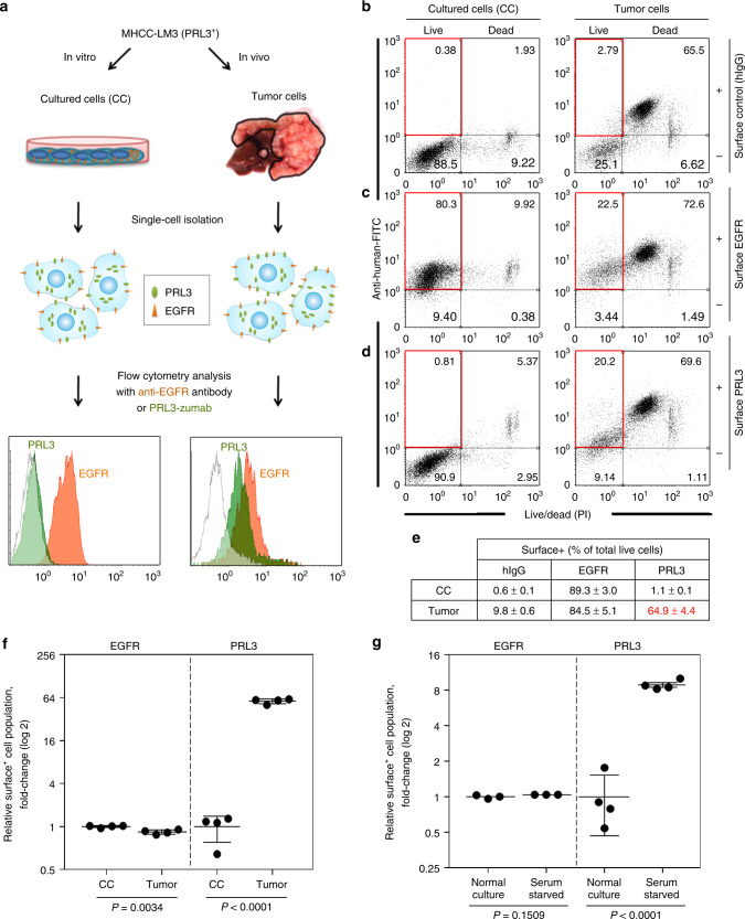
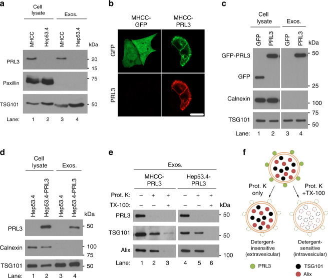
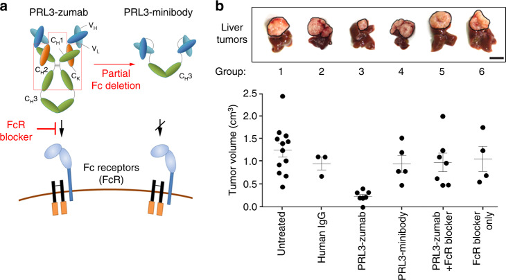
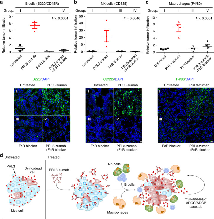
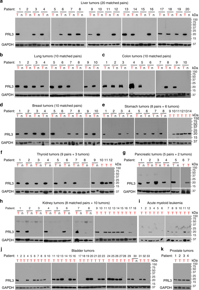
Tumor-specific antibody drugs can serve as cancer therapy with minimal side effects. A humanized antibody, PRL3-zumab, specifically binds to an intracellular oncogenic phosphatase PRL3, which is frequently expressed in several cancers. Here we show that PRL3-zumab specifically inhibits PRL3+ cancer cells in vivo, but not in vitro. PRL3 antigens are detected on the cell surface and outer exosomal membranes, implying an 'inside-out' externalization of PRL3. PRL3-zumab binds to surface PRL3 in a manner consistent with that in classical antibody-dependent cell-mediated cytotoxicity or antibody-dependent cellular phagocytosis tumor elimination pathways, as PRL3-zumab requires an intact Fc region and host FcγII/III receptor engagement to recruit B cells, NK cells and macrophages to PRL3+ tumor microenvironments. PRL3 is overexpressed in 80.6% of 151 fresh-frozen tumor samples across 11 common cancers examined, but not in patient-matched normal tissues, thereby implicating PRL3 as a tumor-associated antigen. Targeting externalized PRL3 antigens with PRL3-zumab may represent a feasible approach for anti-tumor immunotherapy.


Conflict of interest statement

Q.Z. is the founder of Intra-Immu SG Pte Ltd., an Agency of Science, Technology and Research (A*STAR) spin-off company granted licensing rights for the PRL3-zumab IP portfolio. The other authors declare no competing interests.

Figures

Fig. 1
Fig. 1
PRL3-zumab inhibits PRL3+ liver tumors in vivo but not cancer cells in vitro. a Representative western blot (WB) of PRL3 protein expression in human (lanes 1–6) and murine (lanes 7 and 8) liver cancer cells. Glyceraldehyde 3-phosphate dehydrogenase (GAPDH) served as a loading control. Asterisks indicate cell lines that rapidly generate orthotopic liver tumors within 5 weeks. b Outline of orthotopic “seed and soil” liver tumor model for treatments. ce Mean volumes at the end of the experiment in treated (filled squares) and untreated (filled triangles) groups of mice bearing PRL3+ MHCC-LM3 tumors (n = 10 mice per group; c), PRL3 Hep53.4 tumors (n = 3 mice per group; d), and Hep53.4-PRL3 tumors (n = 6 mice per group; e). The mean value was calculated by the Student’s t test (mean ± s.e.m.). P values between treatment pairs as indicated. Lower panels, representative liver tumors at the end of experiment. Scale bar, 10 mm. fh The viabilities of MHCC-LM3 cells (f), Hep53.4 cells (g), and Hep53.4-PRL3 cells (h) cultured for 48 h with PBS control (filled squares), 5 µg mL−1 PRL3-zumab (filled upright triangles), 50 µg mL−1 PRL3-zumab (filled inverted triangles), 2 µg mL−1 cisplatin (filled diamonds), or 10 µg mL−1 cisplatin (filled circles) were evaluated by an MTS (3-(4,5-dimethylthiazol-2-yl)−5-(3-carboxymethoxyphenyl)−2-(4-sulfophenyl)-2H-tetrazolium) assay. The mean value was calculated by the Student’s t test (mean ± s.e.m., n = 3 biologically independent samples each). *P< 0.05, **P < 0.01, NS, not significant, as compared between treatment and control group for each cell line. Source data are provided as a Source Data file
Fig. 2
Fig. 2
Intracellular PRL3 is externalized for PRL3-zumab binding. a Methodology for cell surface analysis of MHCC-LM3 cultured cells (CC) and tumor cells. bd “Surface” detection of nonspecific control antigens (b), EGFR (c), or PRL3 (d) were detected by fluorescence-activated cell sorting (FACS) analysis using polyclonal human hIgG, anti-EGFR antibody (cetuximab), or anti-PRL3 antibody (PRL3-zumab), respectively. Representative FACS profiles from four biological replicates are shown. e Mean percentage ± s.d. of surface positive (surface+) live cells for each antigen were calculated by dividing the surface antigen-positive live cells (upper left quadrant) by total live cells (sum of both upper and lower left quadrants) in bd. f Background-corrected values from e were normalized to CC surface expression levels for EGFR (filled circles) and PRL3 (filled squares). The mean fold change was calculated by the Student’s t test (mean ± s.d., n = 4 biologically independent samples). P values as indicated for each antigen. g Background-corrected values of MHCC-LM3 cells cultured under “Normal” vs. “Serum-starved” conditions for 72 h were normalized to “Normal” surface+ cell percentages for each antigen. The mean fold-change was calculated by the Student’s t test (mean ± s.d.) for EGFR (filled circles; n = 3 independent samples) and PRL3 (filled squares; n = 4 independent samples). P values as indicated for each antigen. Source data are provided as a Source Data file
Fig. 3
Fig. 3
PRL3 is expressed on the surface of exosomes from PRL3+ cancer cells. a Representative PRL3 western blot (WB) in purified exosomes from MHCC-LM3 and Hep53.4 liver cancer cells cultured for 24 h in serum-free media. Paxillin served as a negative marker for exosomes, whereas TSG101 is a positive exosomal marker. b Immunofluorescence analysis of MHCC-LM3 cells transiently transfected with green fluorescence protein (GFP) or GFP-PRL3 to reveal plasma membrane enrichment of PRL3. Scale bar, 20 µm. c Representative PRL3 WB in purified exosomes from cells in b after culturing for 24 h in serum-free media. Calnexin served as a negative marker for exosomes. d Purified exosomes from Hep53.4 and Hep53.4-PRL3 were analyzed as in c. e Purified exosomes from cells in c, d were assayed for sensitivity of exosomal PRL3 to proteinase K (Prot. K) digestion in the absence or presence of 1% Triton X-100 detergent. TG101 and Alix are intravesicular exosomal proteins. f Proposed model of the localization of PRL3, TSG101, and Alix in exosomes, based on Prot. K susceptibility observed in e. Source data are provided as a Source Data file
Fig. 4
Fig. 4
PRL3-zumab eliminates tumors in an Fc- and FcR-dependent manner. a Cartoon depicting domain architecture of PRL3-zumab (intact Fc) vs. PRL3-minibody (truncated Fc lacking CH1 and CH2 domains) and their ability to engage Fc receptors (FcR) on host immune cells. The anti-CD16/32 FcR blocker antibody prevents IgG from binding murine FcγII/III receptors. b Both Fc region of PRL3-zumab and FcR binding are required for anti-tumor effects of PRL3-zumab. Upper panel, representative images of livers from each treatment group at day 35 (5-week endpoint). Tumor areas are framed with black lines. Scale bar, 10 mm. Lower panel, tumor volumes in each group. The mean tumor volumes were calculated using one-way analysis of variance (ANOVA) (mean ± s.e.m.) for independent groups of untreated (n = 12), human IgG-treated (n = 3), PRL3-zumab-treated (n = 6), PRL3-minibody-treated (n = 5), PRL3-zumab + FcR blocked-treated (n = 7), and FcR blocker-treated (n = 4) mice. Source data are provided as a Source Data file
Fig. 5
Fig. 5
PRL3-zumab recruits immune cells to tumor sites in an FcR-dependent manner. ac Orthotopic MHCC-LM3 liver tumor tissue cryo-sections from various groups of mice: I) untreated (filled black squares), II) PRL3-zumab treatment (filled red triangles), III) FcR blocker treatment (filled black triangles), and IV) PRL3-zumab plus Fc blocker combination treatment (filled black diamonds) were analyzed by immunofluorescence with antibodies against B220/CD45R (B cells; a), CD335 (NK cells; b), or F4/80 (macrophages; c) and scored for relative tumor infiltration. The mean relative infiltration was calculated using one-way analysis of variance (ANOVA) (mean ± s.e.m., n = 4 independent samples). Lower panels, representative immunofluorescence results for each antibody set. Scale bar, 200 µm. d Proposed mechanism of action of PRL3-zumab. Externalized PRL3 antigens are recognized by PRL3-zumab, which then recruits NK cells, B cells, and Ly-6C+F4/80+ macrophages for tumor killing and leakage of additional PRL3 antigens (“kill-and-leak” cycle) triggering an anti-tumor ADCC/ADCP cascade. Source data are provided as a Source Data file
Fig. 6
Fig. 6
PRL3 is frequently overexpressed in multiple human cancers. ak Representative full western blot (WB) of PRL3 protein levels in tumor (“T”) tissues and, where available, patient-matched normal (“n”) tissues from a liver, b lung, c colon, d breast, e stomach, f thyroid, g pancreas, h kidney, i acute myeloid leukemia (bone marrow aspirates), j bladder, and k prostate tissues. Relative molecular masses (in kDa) are indicated on the right of each immunoblot. Glyceraldehyde 3-phosphate dehydrogenase (GAPDH) served as a loading control. Source data are provided as a Source Data file

References

    1. Imai K, Takaoka A. Comparing antibody and small-molecule therapies for cancer. Nat. Rev. Cancer. 2006;6:714–727. doi: 10.1038/nrc1913. - DOI - PubMed
    1. Strebhardt K, Ullrich A. Paul Ehrlich’s magic bullet concept: 100 years of progress. Nat. Rev. Cancer. 2008;8:473–480. doi: 10.1038/nrc2394. - DOI - PubMed
    1. Zeng Q, et al. Prenylation-dependent association of protein-tyrosine phosphatases PRL-1, -2, and -3 with the plasma membrane and the early endosome. J. Biol. Chem. 2000;275:21444–21452. doi: 10.1074/jbc.M000453200. - DOI - PubMed
    1. Zeng Q, Hong W, Tan YH. Mouse PRL-2 and PRL-3, two potentially prenylated protein tyrosine phosphatases homologous to PRL-1. Biochem. Biophys. Res. Commun. 1998;244:421–427. doi: 10.1006/bbrc.1998.8291. - DOI - PubMed
    1. Bessette DC, Qiu D, Pallen CJ. PRL PTPs: mediators and markers of cancer progression. Cancer Metastas. Rev. 2008;27:231–252. doi: 10.1007/s10555-008-9121-3. - DOI - PubMed

Publication types

MeSH terms