. 2019 Jun 6;10(6):450.
doi: 10.1038/s41419-019-1688-9.

The alteration of RhoA geranylgeranylation and Ras farnesylation breaks the integrity of the blood-testis barrier and results in hypospermatogenesis

Affiliations

The alteration of RhoA geranylgeranylation and Ras farnesylation breaks the integrity of the blood-testis barrier and results in hypospermatogenesis

Ruilou Zhu et al. Cell Death Dis. .

Abstract

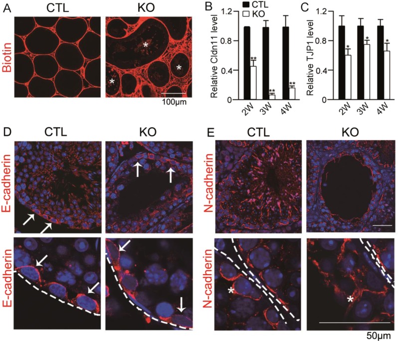
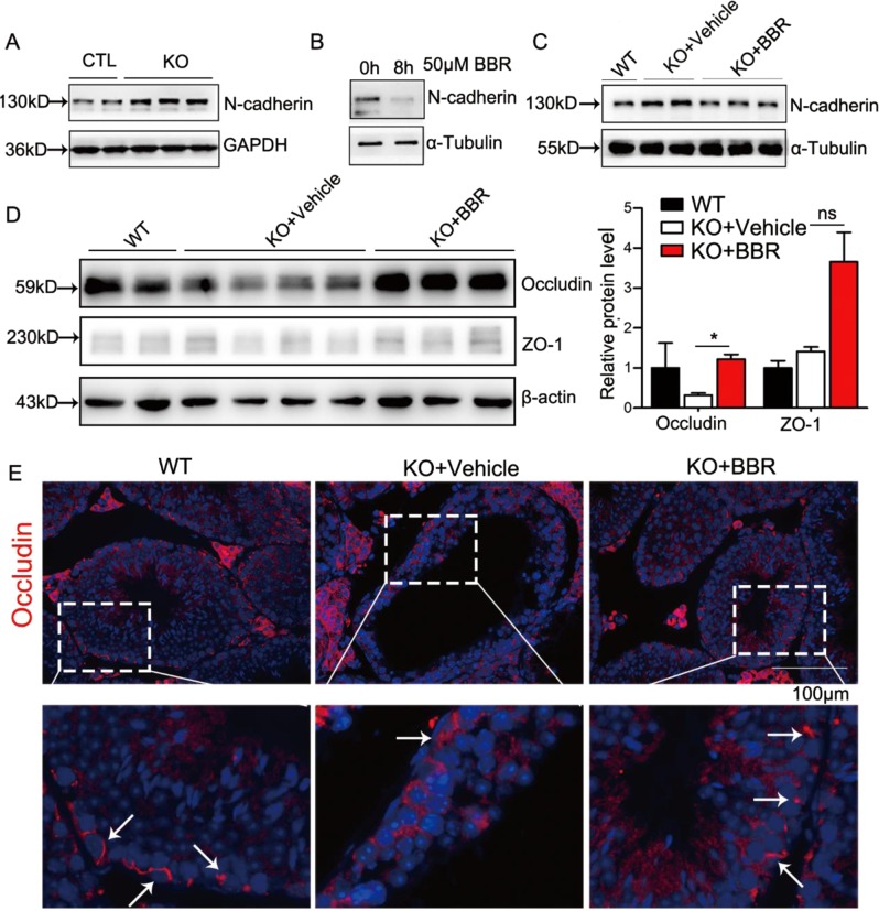
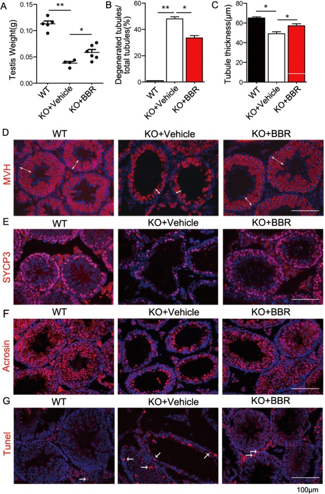
Non-obstructive azoospermia (NOA) severely affects male infertility, however, the deep mechanisms of this disease are rarely interpreted. In this study, we find that undifferentiated spermatogonial stem cells (SSCs) still exist in the basal compartment of the seminiferous tubules and the blood-testis barrier (BTB) formed by the interaction of neighbor Sertoli cells (SCs) is incomplete in NOA patients with spermatogenic maturation arrest. The adhesions between SCs and germ cells (GCs) are also broken in NOA patients. Meanwhile, the expression level of geranylgeranyl diphosphate synthase (Ggpps), a key enzyme in mevalonate metabolic pathway, is lower in NOA patients than that in obstructive azoospermia (OA) patients. After Ggpps deletion specifically in SCs, the mice are infertile and the phenotype of the SC-Ggpps-/- mice is similar to the NOA patients, where the BTB and the SC-GC adhesions are severely destroyed. Although SSCs are still found in the basal compartment of the seminiferous tubules, fewer mature spermatocyte and spermatid are found in SC-Ggpps-/- mice. Further examination suggests that the defect is mediated by the aberrant protein isoprenylation of RhoA and Ras family after Ggpps deletion. The exciting finding is that when the knockout mice are injected with berberine, the abnormal cell adhesions are ameliorated and spermatogenesis is partially restored. Our data suggest that the reconstruction of disrupted BTB is an effective treatment strategy for NOA patients with spermatogenic maturation arrest and hypospermatogenesis.


Conflict of interest statement

The authors declare that they have no conflicts of interest.

Figures

Fig. 1
Fig. 1. Disruption of the blood–testis barrier leads to spermatogenesis arrest in NOA patients.
a, b Immunofluorescence of GC marker MVH and undifferentiated SSC marker Plzf in OA and NOA patients. The arrows indicate the positive staining. c, d Immunofluorescence staining of TJ-associated protein ZO-1 and adherens junction protein N-cadherin in OA and NOA patients. e Immunohistochemistry and density of the positive staining of GGPPS in OA and NOA patients. f Relative mRNA level of GGPPS in OA and NOA patients. n = 18 of NOA patients and n = 5 of OA patients to be performed for immunofluorescence and immunohistochemistry, respectively. n = 7 of NOA patients and n = 3 of OA patients to be performed for qRT-PCR, respectively. Data are presented as the mean ± SEM. *p < 0.05; **p < 0.01, n ≥ 3. Scale bar: 100 μm
Fig. 2
Fig. 2. Ggpps deletion results in GC loss in the adluminal compartment while not in the basal compartment.
a H&E staining of mice testis in CTL and KO mice at 3 weeks old. b Immunofluorescence of GC marker MVH in CTL and KO mice testis at 3 weeks old. c Co-immunofluorescence of spermatocyte marker Sycp3 and TJ-associated protein ZO-1 at 2 weeks old. d Co-immunofluorescence of the undifferentiated SSC marker Plzf and proliferation marker Ki67 at 2 weeks old. The white arrow indicates the co-staining of Plzf and Ki67. e Tunel assay in CTL and KO mice testis. The red arrow indicates the Tunel-positive staining. f Immunofluorescence of adherens junction protein n-cadherin in CTL and KO mice testis. The asterisk indicates the distribution of N-cadherin in apical cytoplasm of SCs. n ≥ 3
Fig. 3
Fig. 3. Ggpps deletion in SC leads to the destruction of BTB and spermatocyte–SC adhesion.
a Biotin distribution in CTL and KO mice testis after biotin injection at 4 weeks old. The asterisk indicates the biotin penetrating into seminiferous tubules. b, c RT-qPCR analysis of tight junction protein: claudin11 and TJP1 during BTB establishment. d, e Immunofluorescence of adherens junction protein E-cadherin and N-cadherin in CTL and KO mice testis. The dashed line indicates the bottom of seminiferous tubules. The white arrow indicates the distribution of E-cadherin and N-cadherin in testis. Data are presented as the mean ± SEM. *p < 0.05, **p < 0.01, n ≥ 3
Fig. 4
Fig. 4. Ggpps affects the BTB and cell adhesion via the regulation of protein isoprenylation of small G-protein.
a, b Immunoblot with Rho family members Cdc42 and RhoA of hydrophobic proteins (down) and hydrophilic proteins (up) in CTL and KO mice testis. c Immunoblot with the active RhoA form RhoA-GTP in CTL and KO mice testis. d Immunoblot with Pan-Ras, H-Ras, and K-Ras in hydrophobic proteins (down) and hydrophilic (up) proteins of primary SC. e Immunoblot with p-ERK and T-ERK of primary SC in control and KO mice relative to α-tubulin. f Immunoblot with p-ERK and T-ERK after FTI treatment of primary SC in control and KO mice relative to α-tubulin. gj Relative mRNA level of tight junction proteins after FTI treatment. Data are presented as the mean ± SEM. *p < 0.05; **p < 0.01, n ≥ 3
Fig. 5
Fig. 5. Berberine treatment could ameliorate the damaged tight junction.
a Immunoblot with n-cadherin in CTL and KO mice testis relative to GAPDH. b Immunoblot with N-cadherin of primary SC in KO mice after berberine treatment relative to α-Tubulin. c Immunoblot with N-cadherin in WT and KO mice testis after berberine treatment relative to α-tubulin. d Immunoblot and statistical analysis of tight junction proteins occludin and ZO-1 in WT and KO mice testis after berberine treatment relative to β-actin. e Immunofluorescence with occludin in WT and KO mice testis after berberine treatment. Data are presented as the mean ± SEM. *p < 0.05; **p < 0.01, n ≥ 3. Scale bar: 100 μm
Fig. 6
Fig. 6. Berberine restores spermatogenesis via ameliorating the damaged tight junction.
a, b Analysis of testis weights and degenerated tubules in KO mice testis after berberine treatment relative to WT mice. c, d The epithelium thickness and immunofluorescence with GC marker MVH in WT and KO mice testis after berberine treatment. White double arrow indicates the epithelium thickness. e, f Immunofluorescence with spermatocyte marker Sycp3 and round spermatid marker acrosin in WT and KO mice after berberine treatment. g Tunel assay in WT and KO mice after berberine treatment. Data are presented as the mean ± SEM. *p < 0.05, **p < 0.01, n ≥ 3

References

    1. Nakagawa T, Nabeshima Y, Yoshida S. Functional identification of the actual and potential stem cell compartments in mouse spermatogenesis. Dev. Cell. 2007;12:195–206. doi: 10.1016/j.devcel.2007.01.002. - DOI - PubMed
    1. Oatley JM, Brinster RL. The germline stem cell niche unit in mammalian testes. Physiol. Rev. 2012;92:577–595. doi: 10.1152/physrev.00025.2011. - DOI - PMC - PubMed
    1. Dym M, Fawcett DW. The blood-testis barrier in the rat and the physiological compartmentation of the seminiferous epithelium. Biol. Reprod. 1970;3:308–326. doi: 10.1093/biolreprod/3.3.308. - DOI - PubMed
    1. Cheng CY, Mruk DD. The blood–testis barrier and its implications for male contraception. Pharmacol Rev. 2012;64:16–64. doi: 10.1124/pr.110.002790. - DOI - PMC - PubMed
    1. Enders GC, Millette CF. Pachytene spermatocyte and round spermatid binding to Sertoli cells in vitro. J. Cell Sci. 1988;90(Part 1):105–114. - PubMed

Publication types

MeSH terms

Supplementary concepts

LinkOut - more resources