

The human glomerular endothelial cells are potent pro-inflammatory contributors in an in vitro model of lupus nephritis
- PMID: 31171837
- PMCID: PMC6554346
- DOI: 10.1038/s41598-019-44868-y
The human glomerular endothelial cells are potent pro-inflammatory contributors in an in vitro model of lupus nephritis
Abstract
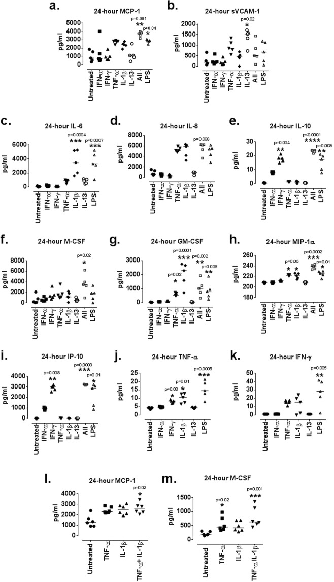
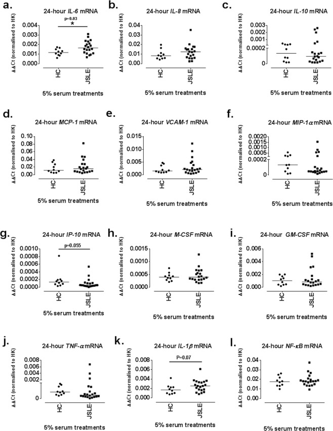
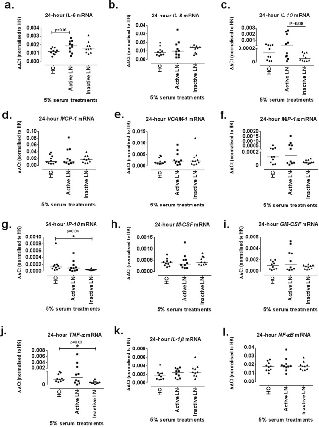
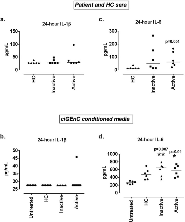
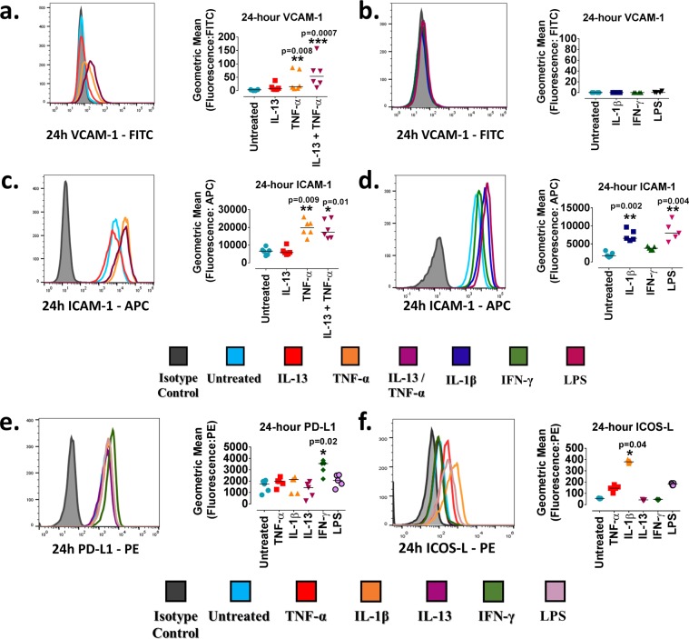
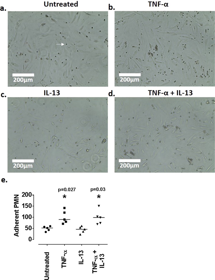
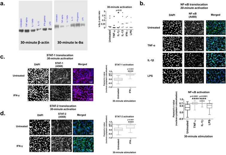
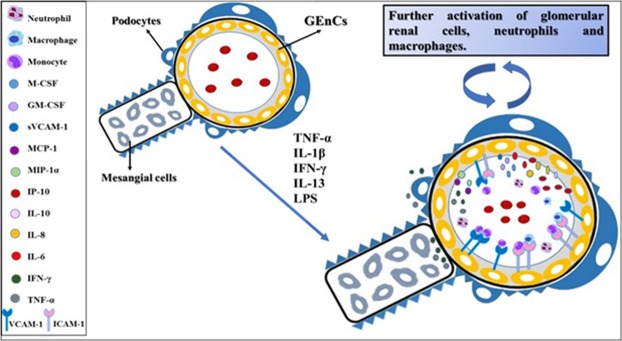
Juvenile-onset lupus nephritis (LN) affects up to 80% of juvenile-onset systemic lupus erythematosus patients (JSLE). As the exact role of human renal glomerular endothelial cells (GEnCs) in LN has not been fully elucidated, the aim of this study was to investigate their involvement in LN. Conditionally immortalised human GEnCs (ciGEnCs) were treated with pro-inflammatory cytokines known to be involved in LN pathogenesis and also with LPS. Secretion and surface expression of pro-inflammatory proteins was quantified via ELISA and flow cytometry. NF-κΒ and STAT-1 activation was investigated via immunofluorescence. Serum samples from JSLE patients and from healthy controls were used to treat ciGEnCs to determine via qRT-PCR potential changes in the mRNA levels of pro-inflammatory genes. Our results identified TNF-α, IL-1β, IL-13, IFN-γ and LPS as robust in vitro stimuli of ciGEnCs. Each of them led to significantly increased production of different pro-inflammatory proteins, including; IL-6, IL-10, MCP-1, sVCAM-1, MIP-1α, IP-10, GM-CSF, M-CSF, TNF-α, IFN-γ, VCAM-1, ICAM-1, PD-L1 and ICOS-L. TNF-α and IL-1β were shown to activate NF-κB, whilst IFN-γ activated STAT-1. JSLE patient serum promoted IL-6 and IL-1β mRNA expression. In conclusion, our in vitro model provides evidence that human GEnCs play a pivotal role in LN-associated inflammatory process.
Conflict of interest statement
The authors declare no competing interests.
Figures
Similar articles
-
In situ glomerular expression of activated NF-kappaB in human lupus nephritis and other non-proliferative proteinuric glomerulopathy.Virchows Arch. 2006 Feb;448(2):172-83. doi: 10.1007/s00428-005-0061-9. Epub 2005 Oct 5. Virchows Arch. 2006. PMID: 16205945
-
Podocytes contribute, and respond, to the inflammatory environment in lupus nephritis.Am J Physiol Renal Physiol. 2018 Dec 1;315(6):F1683-F1694. doi: 10.1152/ajprenal.00512.2017. Epub 2018 Sep 12. Am J Physiol Renal Physiol. 2018. PMID: 30207171 Free PMC article.
-
CD8+iTregs attenuate glomerular endothelial cell injury in lupus-prone mice through blocking the activation of p38 MAPK and NF-κB.Mol Immunol. 2018 Nov;103:133-143. doi: 10.1016/j.molimm.2018.09.006. Epub 2018 Sep 27. Mol Immunol. 2018. PMID: 30268079
-
A novel potential target of IL-35-regulated JAK/STAT signaling pathway in lupus nephritis.Clin Transl Med. 2021 Feb;11(2):e309. doi: 10.1002/ctm2.309. Clin Transl Med. 2021. PMID: 33634995 Free PMC article.
-
Proteolysis and inflammation of the kidney glomerulus.Cell Tissue Res. 2021 Aug;385(2):489-500. doi: 10.1007/s00441-021-03433-8. Epub 2021 Apr 17. Cell Tissue Res. 2021. PMID: 33864499 Free PMC article. Review.
Cited by
-
Induction of PD-1 and CD44 in CD4+ T cells by circulatory extracellular vesicles from severe dengue patients drives endothelial damage via the NF-kB signaling pathway.J Virol. 2025 Feb 25;99(2):e0186124. doi: 10.1128/jvi.01861-24. Epub 2024 Dec 31. J Virol. 2025. PMID: 39745465 Free PMC article.
-
Lupus serum induces inflammatory interaction with neutrophils in human glomerular endothelial cells.Lupus Sci Med. 2020 Oct;7(1):e000418. doi: 10.1136/lupus-2020-000418. Lupus Sci Med. 2020. PMID: 33037079 Free PMC article.
-
Endothelial function and endothelial progenitor cells in systemic lupus erythematosus.Nat Rev Rheumatol. 2022 May;18(5):286-300. doi: 10.1038/s41584-022-00770-y. Epub 2022 Apr 7. Nat Rev Rheumatol. 2022. PMID: 35393604 Review.
-
Endothelial cell activation and glycocalyx shedding - potential as biomarkers in patients with lupus nephritis.Front Immunol. 2023 Oct 3;14:1251876. doi: 10.3389/fimmu.2023.1251876. eCollection 2023. Front Immunol. 2023. PMID: 37854589 Free PMC article. Review.
-
Role of the transcription factor Fli-1 on the CXCL10/CXCR3 Axis.Front Immunol. 2023 Sep 15;14:1219279. doi: 10.3389/fimmu.2023.1219279. eCollection 2023. Front Immunol. 2023. PMID: 37790939 Free PMC article.
References
Publication types
MeSH terms
Substances
Grants and funding
LinkOut - more resources
Full Text Sources
Research Materials
Miscellaneous
