. 2019 Jun 4:19:155.
doi: 10.1186/s12935-019-0867-1. eCollection 2019.

Systematically profiling the expression of eIF3 subunits in glioma reveals the expression of eIF3i has prognostic value in IDH-mutant lower grade glioma

Affiliations

Systematically profiling the expression of eIF3 subunits in glioma reveals the expression of eIF3i has prognostic value in IDH-mutant lower grade glioma

Rui-Chao Chai et al. Cancer Cell Int. .

Abstract

Background: Abnormal expression of the eukaryotic initiation factor 3 (eIF3) subunits plays critical roles in tumorigenesis and progression, and also has potential prognostic value in cancers. However, the expression and clinical implications of eIF3 subunits in glioma remain unknown.

Methods: Expression data of eIF3 for patients with gliomas were obtained from the Chinese Glioma Genome Atlas (CGGA) (n = 272) and The Cancer Genome Atlas (TCGA) (n = 595). Cox regression, the receiver operating characteristic (ROC) curves and Kaplan-Meier analysis were used to study the prognostic value. Gene oncology (GO) and gene set enrichment analysis (GSEA) were utilized for functional prediction.

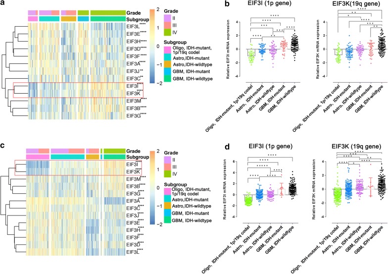
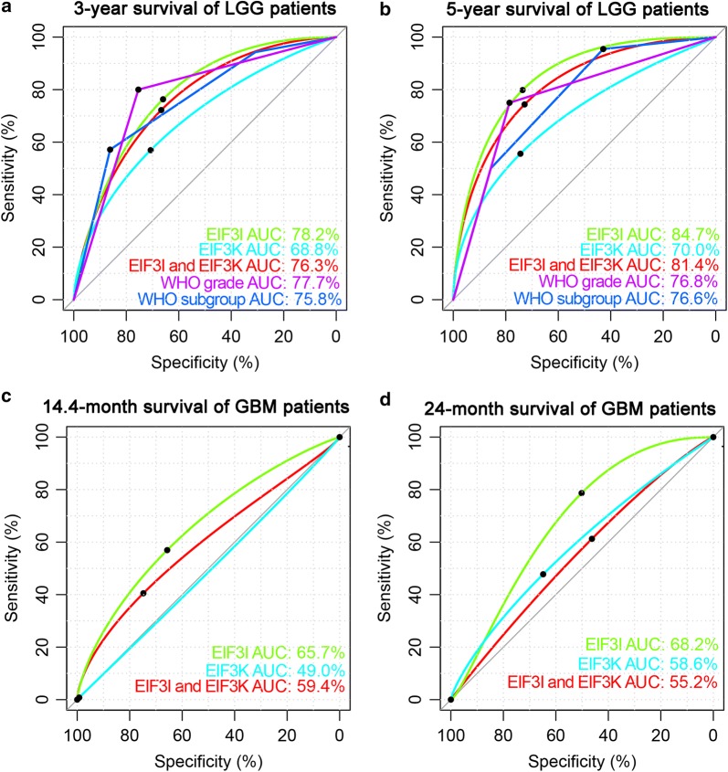
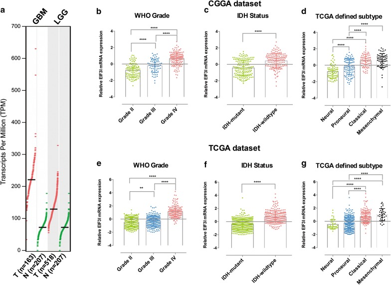
Results: In both the CGGA and TCGA datasets, the expression levels of eIF3d, eIF3e, eIF3f, eIF3h and eIF3l highly were associated with the IDH mutant status of gliomas. The expression of eIF3b, eIF3i, eIF3k and eIF3m was increased with the tumor grade, and was associated with poorer overall survival [All Hazard ratio (HR) > 1 and P < 0.05]. By contrast, the expression of eIF3a and eIF3l was decreased in higher grade gliomas and was associated with better overall survival (Both HR < 1 and P < 0.05). Importantly, the expression of eIF3i (located on chromosome 1p) and eIF3k (Located on chromosome 19q) were the two highest risk factors in both the CGGA [eIF3i HR = 2.068 (1.425-3.000); eIF3k HR = 1.737 (1.166-2.588)] and TCGA [eIF3i HR = 1.841 (1.642-2.064); eIF3k HR = 1.521 (1.340-1.726)] databases. Among eIF3i, eIF3k alone or in combination, the expression of eIF3i was the more robust in stratifying the survival of glioma in various pathological subgroups. The expression of eIF3i was an independent prognostic factor in IDH-mutant lower grade glioma (LGG) and could also predict the 1p/19q codeletion status of IDH-mutant LGG. Finally, GO and GSEA analysis showed that the elevated expression of eIF3i was significantly correlated with the biological processes of cell proliferation, mRNA processing, translation, T cell receptor signaling, NF-κB signaling and others.

Conclusions: Our study reveals the expression alterations during glioma progression, and highlights the prognostic value of eIF3i in IDH-mutant LGG.

Keywords: 1p/19q codeletion; Biomarker; Glioma; Prognosis; eIF3.


Conflict of interest statement

Competing interestsThe authors declare that they have no competing interests.

Figures

Fig. 1
Fig. 1
The expression of eIF3 subunits in gliomas with different pathological features. Heatmaps showing the expression levels of thirteen eIF3 subunits in gliomas with different pathological features according to WHO 2016 integrated diagnosis from the CGGA (a) and TCGA (c) datasets. Statistic data showing the expression levels of eIF3i (locate on chromosome 1p) and eIF3k (located on chromosome 19q) in gliomas with different pathological features according to WHO 2016 integrated diagnosis from the CGGA (b) and TCGA (d) datasets. *P < 0.05, **P < 0.01, *** P < 0.001 and ****P < 0.0001
Fig. 2
Fig. 2
The predict efficiencies of eIF3i and eIF3k in gliomas. ROC curves showed the predictive efficiencies of eIF3i, eIF3k, combination of eIF3i and eIF3k, WHO grade and subgroups of WHO 2016 integrated diagnosis (Oligodendroglioma, IDH-mutant, 1p/19q codeletion; Astrocytoma, IDH-mutant; Astrocytoma, IDH-wildtype) on 3-year (a) and 5-year survival (b) in LGG of CGGA dataset. ROC curves showed the predictive efficiencies of eIF3i, eIF3k and combination of eIF3i and eIF3k on 14.4-month (c) and 24-month survival (d) in GBM of CGGA dataset
Fig. 3
Fig. 3
The expression of eIF3i in normal brain tissue and gliomas with different clinicopathological features. a The expression profiles of eIF3i mRNA in each GBM, LGG, normal brain tissues were represented. Quantification data shows the expression levels of eIF3i in gliomas from the CGGA (bd) and TCGA (eg) datasets stratified by the WHO grade (b, e), IDH status (c, f) TCGA defined subtype (d, g). **P < 0.01 and ****P < 0.0001
Fig. 4
Fig. 4
Prognostic value of eIF3i expression in stratified LGG, especially in IDH-mutant LGG. ah Kaplan–Meier overall survival curves for patients stratified by the respective median expression of eIF3i in the CGGA (ad) and TCGA (eh) datasets with astrocytoma, IDH-wildtype (a, e), all IDH-mutant LGGs (b, f), oligodendroglioma, IDH-mutant, 1p/19q codel (c, g) and astrocytoma, IDH-mutant (d, h), respectively. i, j ROC curves showed the predictive efficiencies of eIF3i, WHO grade (II and III) and 1p/19q codeletion status on 3-year and 5-year survival in TCGA (I) and CGGA (J) datasets. k Univariate and multivariate Cox regression analyses of the association between clinicopathological factors (including the eIF3i expression) and overall survival of patients in the CGGA and TCGA datasets
Fig. 5
Fig. 5
Functional annotation for genes significantly correlated to eIF3i expression in IDH-mutant LGG. Biological processes of genes that correlated to eIF3i expression in CGGA (a) and TCGA (b) datasets. c GSEA revealed enriched GO terms positively correlated to EIF3I expression in CGGA dataset

References

    1. Jiang T, Mao Y, Ma W, Mao Q, You Y, Yang X, Jiang C, Kang C, Li X, Chen L, et al. CGCG clinical practice guidelines for the management of adult diffuse gliomas. Cancer Lett. 2016;375(2):263–273. doi: 10.1016/j.canlet.2016.01.024. - DOI - PubMed
    1. Ostrom QT, Gittleman H, Xu J, Kromer C, Wolinsky Y, Kruchko C, Barnholtz-Sloan JS. CBTRUS statistical report: primary brain and other central nervous system tumors diagnosed in the United States in 2009–2013. Neuro-oncology. 2016;18(suppl_5):v1–v75. doi: 10.1093/neuonc/now207. - DOI - PMC - PubMed
    1. Chai R, Zhang K, Wang K, Li G, Huang R, Zhao Z, Liu Y, Chen J. A novel gene signature based on five glioblastoma stem-like cell relevant genes predicts the survival of primary glioblastoma. J Cancer Res Clin Oncol. 2018;144(3):439–447. doi: 10.1007/s00432-017-2572-6. - DOI - PMC - PubMed
    1. Khasraw M, Lassman AB. Advances in the treatment of malignant gliomas. Curr Oncol Rep. 2010;12(1):26–33. doi: 10.1007/s11912-009-0077-4. - DOI - PubMed
    1. Louis DN, Perry A, Reifenberger G, von Deimling A, Figarella-Branger D, Cavenee WK, Ohgaki H, Wiestler OD, Kleihues P, Ellison DW. The 2016 World Health Organization classification of tumors of the central nervous system: a summary. Acta Neuropathol. 2016;131(6):803–820. doi: 10.1007/s00401-016-1545-1. - DOI - PubMed

LinkOut - more resources