. 2019 Dec;21(4):493-504.
doi: 10.1007/s12017-019-08552-8. Epub 2019 Jun 6.

Measuring Respiration in Isolated Murine Brain Mitochondria: Implications for Mechanistic Stroke Studies

Affiliations

Measuring Respiration in Isolated Murine Brain Mitochondria: Implications for Mechanistic Stroke Studies

Jared A Sperling et al. Neuromolecular Med. 2019 Dec.

Abstract

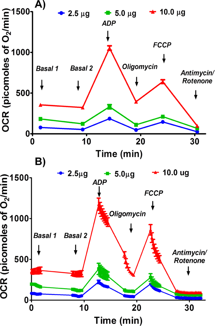
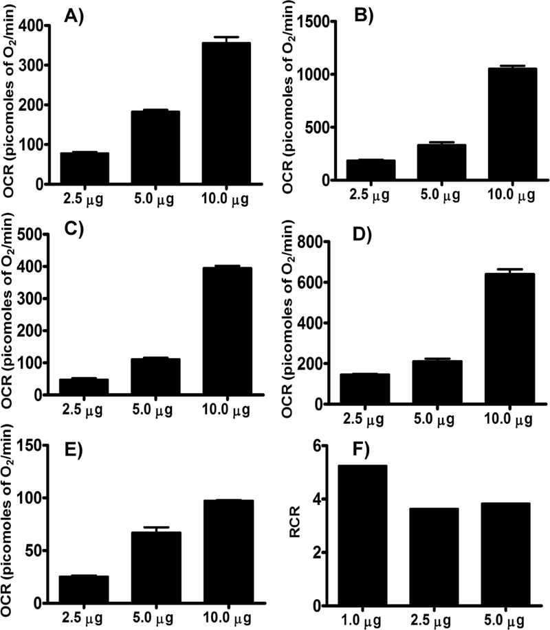
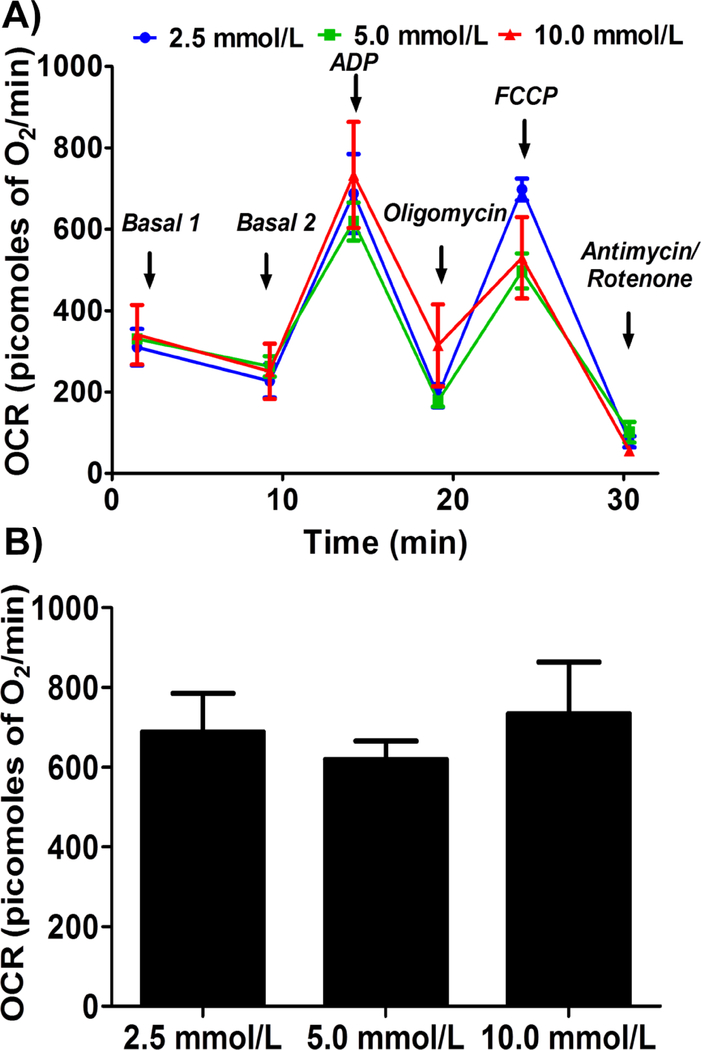
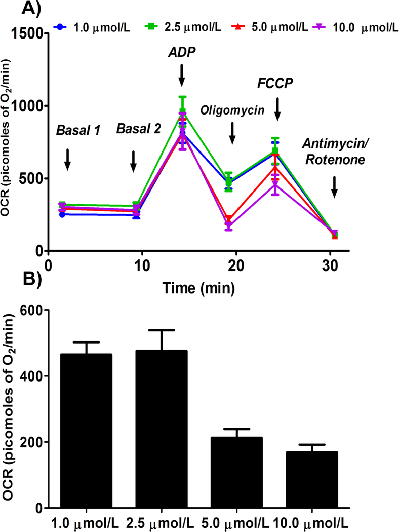
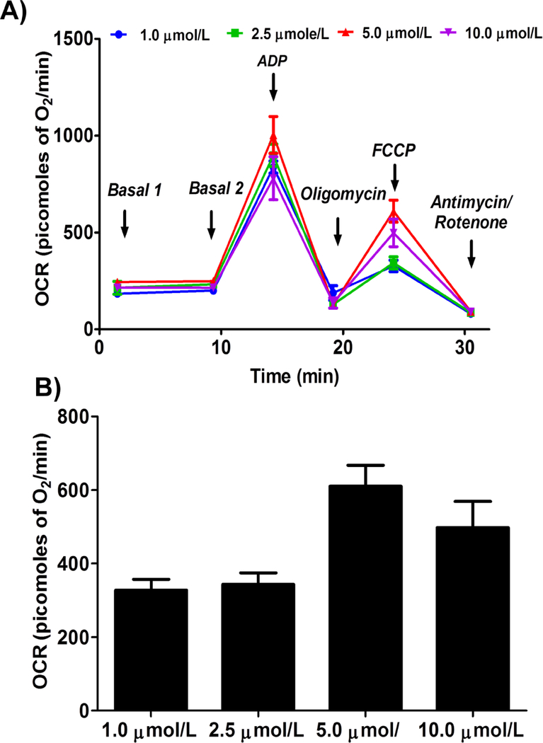
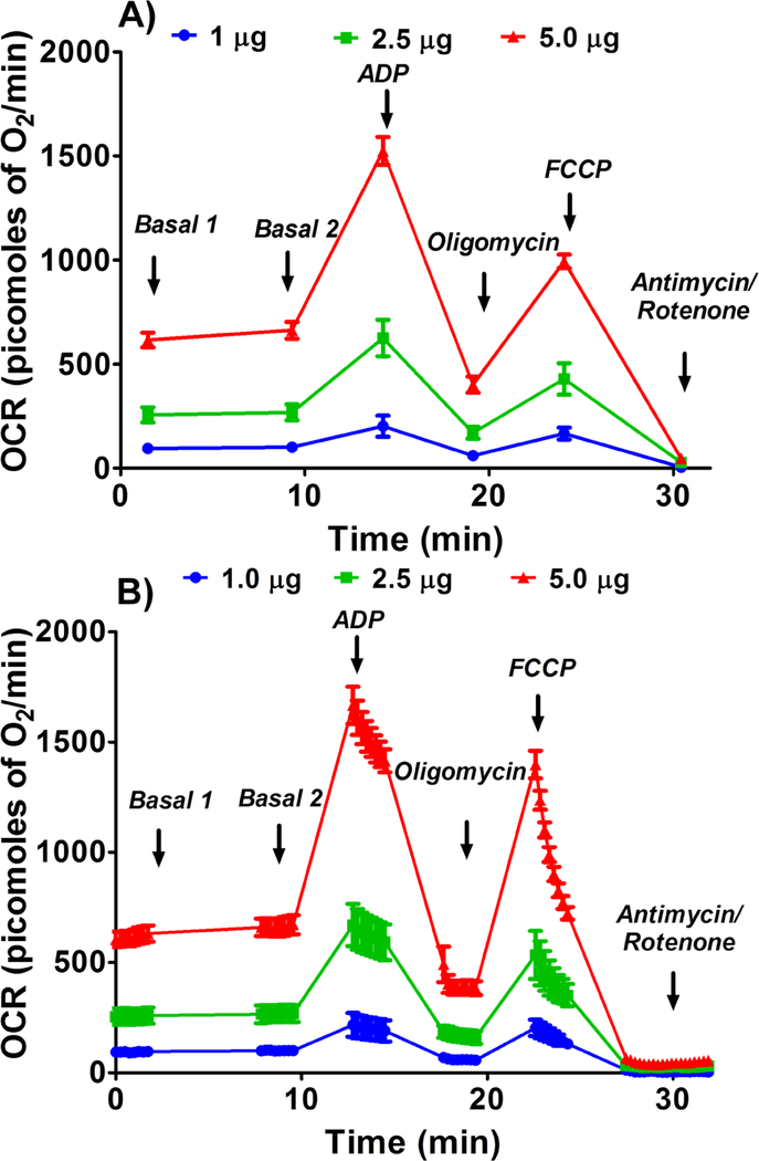
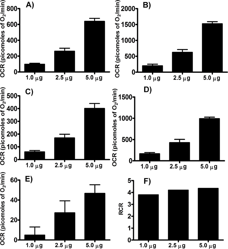
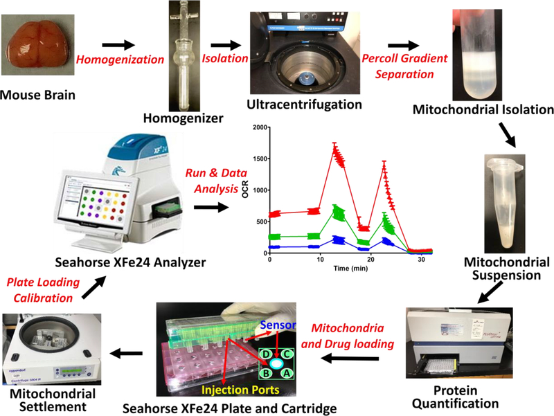
Measuring mitochondrial respiration in brain tissue is very critical in understanding the physiology and pathology of the central nervous system. Particularly, measurement of respiration in isolated mitochondria provides the advantage over the whole cells or tissues as the changes in respiratory function are intrinsic to mitochondrial structures rather than the cellular signaling that regulates mitochondria. Moreover, a high-throughput technique for measuring mitochondrial respiration minimizes the experimental time and the sample-to-sample variation. Here, we provide a detailed protocol for measuring respiration in isolated brain non-synaptosomal mitochondria using Agilent Seahorse XFe24 Analyzer. We optimized the protocol for the amount of mitochondria and concentrations of ADP, oligomycin, and trifluoromethoxy carbonylcyanide phenylhydrazone (FCCP) for measuring respiratory parameters for complex I-mediated respiration. In addition, we measured complex II-mediated respiratory parameters. We observed that 10 µg of mitochondrial protein per well, ADP concentrations ranging between 2.5 and 10 mmol/L along with 5 µmol/L of oligomycin, and 5 µmol/L of FCCP are ideal for measuring the complex I-mediated respiration in isolated mouse brain mitochondria. Furthermore, we determined that 2.5 µg of mitochondrial protein per well is ideal for measuring complex II-mediated respiration. Notably, we provide a discussion of logical analysis of data and how the assay could be utilized to design mechanistic studies for experimental stroke. In conclusion, we provide detailed experimental design for measurement of various respiratory parameters in isolated brain mitochondria utilizing a novel high-throughput technique along with interpretation and analysis of data.

Keywords: Isolated mitochondria; Mitochondrial respiration; Non-synaptosomal mitochondria; Oxygen consumption rate.


Conflict of interest statement

Conflict of interest

The authors declare that they have no conflict of interest.

Figures

Figure 1.
Figure 1.
Optimizing protein concentration for complex I respiratory measurements in isolated mouse brain non-synaptosomal mitochondria using Seahorse XFe24 Analyzer. Isolated mouse (C57BL/6J) non-synaptosomal mitochondria at various concentrations (2.5, 5, and 10 μg protein/well), were treated with ADP (5 mmol/L), oligomycin (5 μmol/L), FCCP (5 μmol/L), and antimycin (10 μmol/L)/rotenone (2 μmol/L) combination in the presence of complex 1 substrates (pyruvate, 10 mmol/L and malate, 2 mmol/L) and oxygen consumption rate (OCR) was measured. A. Representative plot of mid-point OCR data. B. Representative plot of point-to-point OCR. Data expressed as mean ± SEM (n = 5 wells/group).
Figure 2.
Figure 2.
Optimizing protein concentration for complex I respiratory measurements in isolated mouse brain non-synaptosomal mitochondria using Seahorse XFe24 Analyzer. Isolated mouse (C57BL/6J) non-synaptosomal mitochondria at various concentrations (1, 2.5, 5, and 10 μg protein/well), were treated with ADP (5 mmol/L), oligomycin (5 μmol/L), FCCP (5 μmol/L), and antimycin (10 μmol/L)/rotenone (2 μmol/L) combination in the presence of complex 1 substrates (pyruvate, 10 mmol/L and malate, 2 mmol/L) and oxygen consumption rate (OCR) was measured. A. State II or basal respiration. B. State III respiration. C. State IVo respiration. D. State IIIu respiration. E. Antimycin/rotenone. F. Respiratory control ratio (state III/IVo). Data expressed as mean ± SEM (n = 5 wells/group)
Figure 3.
Figure 3.
Optimizing ADP concentration for measuring state III respiration in isolated mouse brain non-synaptosomal mitochondria using Seahorse XFe24 Analyzer.. Isolated mouse (C57BL/6J) non-synaptosomal mitochondria (10 μg protein/well) were treated with various concentrations of ADP (2.5, 5 and 10 mmol/L), oligomycin (5 μmol/L), FCCP (5 μmol/L), and antimycin (10 μmol/L)/rotenone (2 μmol/L) combination in the presence of complex 1 substrates (pyruvate, 10 mmol/L and malate, 2 mmol/L) and oxygen consumption rate (OCR) was measured. A. Representative plot of mid-point OCR data. B. State III respiration. Data expressed as mean ± SEM (n = 5 wells/group)
Figure 4.
Figure 4.
Optimizing oligomycin concentration for measuring state IVo respiration in isolated mouse brain non-synaptosomal mitochondria using Seahorse XFe24 Analyzer. Isolated mouse (C57BL/6J) non-synaptosomal mitochondria (10 μg protein/well) were treated with ADP (10 mmol/L), various doses of oligomycin (1, 2.5, 5 and 10 μmol/L), FCCP (5 μmol/L), and antimycin (10 μmol/L)/rotenone (2 μmol/L) combination in the presence of complex 1 substrates (pyruvate, 10 mmol/L and malate, 2 mmol/L) and oxygen consumption rate (OCR) was measured. A. Representative plot of mid-point OCR data. B. State IVo respiration. Data expressed as mean ± SEM (n = 5 wells/group)
Figure 5.
Figure 5.
Optimizing FCCP (trifluoromethoxy carbonylcyanide phenylhydrazone) concentration for measuring state IIIu (state III uncoupled) respiration in isolated mouse brain non-synaptosomal mitochondria using Seahorse XFe24 Analyzer. Isolated mouse (C57BL/6J) non-synaptosomal mitochondria (10 μg protein/well) were treated with ADP (10 mmol/L), oligomycin (10 μmol/L), various doses of FCCP (1, 2.5, 5 and 10 μmol/L), and antimycin (10 μmol/L)/rotenone (2 μmol/L) combination in the presence of complex 1 substrates (pyruvate, 10 mmol/L and malate, 2 mmol/L) and oxygen consumption rate (OCR) was measured. A. Representative plot of mid-point OCR data. B. State IIIu respiration. Data expressed as mean ± SEM (n = 5 wells/group)
Figure 6.
Figure 6.
Optimizing protein concentration for Complex II respiratory measurements in isolated mouse brain non-synaptosomal mitochondria using Seahorse XFe24 Analyzer. Isolated mouse (C57BL/6J), non-synaptosomal mitochondria at various concentrations (1, 2.5, and 5 μg protein/well), were treated with ADP (5 mmol/L), oligomycin (5 μmol/L), FCCP (5 μmol/L), and antimycin (10 μmol/L) combination in the presence of complex 2 substrate succinate (10mmol/L) and rotenone (2 μmol/L). Oxygen consumption rates (OCR) were measured. A. Representative plot of mid-point OCR data. B. Representative plot of point-to-point OCR. Data expressed as mean ± SEM (n = 5 wells/group).
Figure 7.
Figure 7.
Optimizing protein concentration for Complex II respiratory measurements in isolated mouse brain non-synaptosomal mitochondria using Seahorse XFe24 Analyzer. Isolated mouse (C57BL/6J), non-synaptosomal mitochondria at various concentrations (1, 2.5, and 5 μg protein/well), were treated with ADP (5 mmol/L), oligomycin (5 μmol/L), FCCP (5 μmol/L), and antimycin (10 μmol/L) combination in the presence of complex 2 substrate succinate (10mmol/L) and rotenone (2 μmol/L). Oxygen consumption rates (OCR) were measured. A. State II or basal respiration. B. State III respiration. C. State IVO respiration. D. State IIIu respiration. E. Antimycin/rotenone. F. Respiratory control ratio (state III/IVo). Data expressed as mean ± SEM (n = 5 wells/group).
Figure 8.
Figure 8.
A flow diagram of the method is provided for the benefit of easy understanding of the protocol.

References

    1. Agostini M, Romeo F, Inoue S, Niklison-Chirou MV, Elia AJ, Dinsdale D, et al. (2016). Metabolic reprogramming during neuronal differentiation. Cell Death Differ, 23(9), 1502–1514, doi: 10.1038/cdd.2016.36. - DOI - PMC - PubMed
    1. Andersen JV, Jakobsen E, Waagepetersen HS, & Aldana BI (2019). Distinct differences in rates of oxygen consumption and ATP synthesis of regionally isolated non-synaptic mouse brain mitochondria. J Neurosci Res, doi: 10.1002/jnr.24371. - DOI - PubMed
    1. Attwell D, & Laughlin SB (2001). An energy budget for signaling in the grey matter of the brain. J Cereb Blood Flow Metab, 21(10), 1133–1145, doi: 10.1097/00004647-200110000-00001. - DOI - PubMed
    1. Back T, Hemmen T, & Schuler OG (2004). Lesion evolution in cerebral ischemia. J Neurol, 251(4), 388–397, doi: 10.1007/s00415-004-0399-y. - DOI - PubMed
    1. Barrientos A, Fontanesi F, & Diaz F (2009). Evaluation of the mitochondrial respiratory chain and oxidative phosphorylation system using polarography and spectrophotometric enzyme assays. Curr Protoc Hum Genet, Chapter 19, Unit19 13, doi: 10.1002/0471142905.hg1903s63. - DOI - PMC - PubMed

Publication types

MeSH terms

LinkOut - more resources