Review
. 2019 Sep;11(5):e1559.
doi: 10.1002/wnan.1559. Epub 2019 Jun 6.

Nanovaccines for cancer immunotherapy

Affiliations
Review

Nanovaccines for cancer immunotherapy

Yu Zhang et al. Wiley Interdiscip Rev Nanomed Nanobiotechnol. 2019 Sep.

Abstract

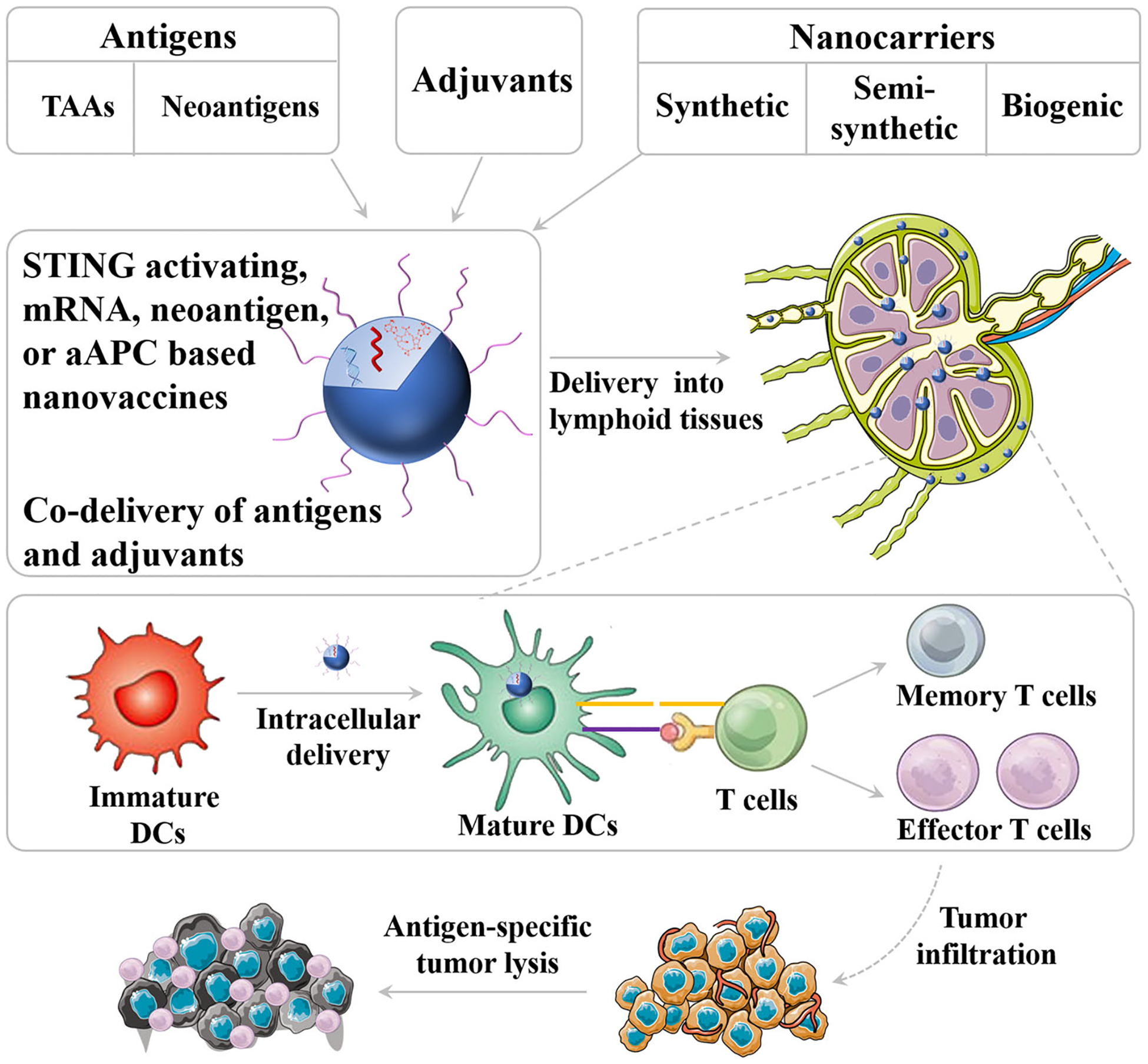
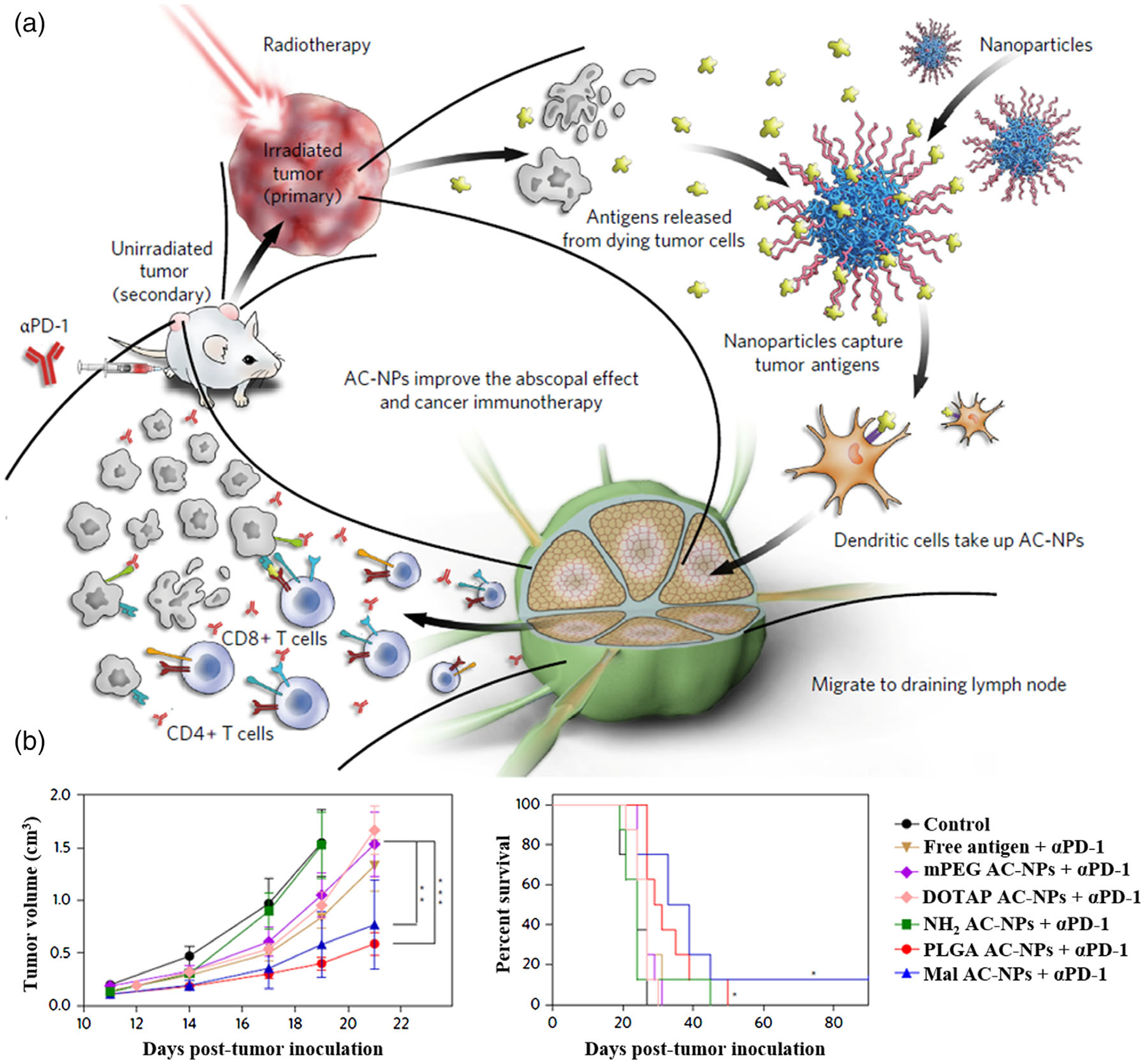
The past few decades have witnessed the booming field of cancer immunotherapy. Cancer therapeutic vaccines, either alone or in combination with other immunotherapies such as adoptive cell therapy or immune checkpoint blockade therapy, are an attractive class of cancer immunotherapeutics. However, cancer vaccines have thus far shown suboptimal efficacy in the clinic. Nanomedicines offer unique opportunities to improve the efficacy of these vaccines. A variety of nanoplatforms have been investigated to deliver molecular or cellular or subcellular vaccines to target lymphoid tissues and cells, thereby promoting the potency and durability of anti-tumor immunity while reducing adverse side effects. In this article, we reviewed the key parameters and features of nanovaccines for cancer immunotherapy; we highlighted recent advances in the development of cancer nanovaccines based on synthetic nanocarriers, biogenic nanocarriers, as well as semi-biogenic nanocarriers; and we summarized newly emerging types of nanovaccines, such as those based on stimulator of interferon genes agonists, cancer neoantigens, mRNA vaccines, as well as artificial antigen-presenting cells. This article is categorized under: Therapeutic Approaches and Drug Discovery > Nanomedicine for Oncologic Disease.

Keywords: STING agonists; cancer immunotherapy; co-delivery; mRNA vaccines; nanocarriers; nanovaccine; neoantigen vaccines.


Conflict of interest statement

CONFLICT OF INTEREST

G.Z. was listed as an inventor for the application of a patent associated with immunomodulatory materials.

Figures

FIGURE 1
FIGURE 1
Schematic depiction of nanovaccine. Nanovaccines can be loaded with both adjuvant and antigens on the surface of nanocarriers or inside nanocarriers. Nanovaccines can efficiently co-deliver adjuvant and tumor antigen to lymphoid tissues and intracellular delivery into antigen-presenting cells (APCs) for efficient induction of antitumor T cell response
FIGURE 2
FIGURE 2
Biogenic nanocarriers of vaccines. (a) Schematic representation of the generation of exosomes. (Reprinted with permission from Jella et al. (2018). Copyright 2018 MDPI Publishing Group) (b) Schematic representation of the generation of engineered antigen-loaded OMVs. (Reprinted with permission from Rosenthal et al. (2014). Copyright 2014 PLOS Publishing Group)
FIGURE 3
FIGURE 3
Cell membrane camouflage-based nanocarriers of vaccines. (a) Schematic representation of the fabrication of cancer cell membrane-coated nanoparticle (CCNP) and the potential applications. (b) CCNPs for the delivery of tumor-associated antigens, and maturation of dendritic cells. (Reprinted with permission from Fang et al. (2014). Copyright 2014 American Chemical Society Publishing Group)
FIGURE 4
FIGURE 4
Endogenous protein-based vaccine carriers. (a) Schematic depiction of albumin/AlbiVax nanocomplexes for efficient vaccine delivery and combination cancer immunotherapy. (b) Albumin/AlbiVax nanocomplexes for melanoma combination immunotherapy. (Reprinted with permission from Zhu, Lynn, et al. (2017). Copyright 2017 Nature Publishing Group)
FIGURE 5
FIGURE 5
Synthetic SNA-based nanovaccines for cancer immunotherapy. (a) Schematic diagram of an SNA platform, which contain a shell of TLR agonist or TLR antagonist. (b) Schematic depiction of proposed mechanism for uptake and TLR interaction between APCs and SNAs. (c–f) Confocal microscopy image showing the Au-SNAs have been internalized. (g) IS-SNAs were tested for their ability to induce NF-κB following incubation with RAW-Blue macrophages overnight. (h) IS-SNAs enhance humoral and cellular immune responses to antigen, independent of core template. (Reprinted with permission from Radovic-Moreno et al. (2015). Copyright 2015 National Academy of Sciences Publishing Group)
FIGURE 6
FIGURE 6
STING agonist-based nanovaccines for cancer immunotherapy. (a) Schematic diagram of NP-MPER and NP-cdGMP (cyclic di-GMP) vaccine. (b) Measurement of the NP-cdGMP vaccine in the blood of mice. (c) Representative flow cytometry plots of CDN fluorescence in APCs 24 hours following s.c. injection. (d) The stability of the NP-cdGMP vaccine in serum. (Reprinted with permission from Hanson et al. (2015). Copyright 2015 American Society for Clinical Investigation Publishing Group)
FIGURE 7
FIGURE 7
Synthetic high-density lipoprotein (sHDL) nanodisc-based personalized cancer therapeutic vaccines. (a) Design of sHDL nanodisc platform for personalized cancer vaccines. (b) Treatment of MC-38 tumor-bearing C57BL/6 mice with sHDL-Ag/CpG vaccines. (Reprinted with permission from Kuai, Ochyl, Bahjat, Schwendeman, & Moon (2017). Copyright 2017 Nature Publishing Group)
FIGURE 8
FIGURE 8
Liposome nanocarrier-based mRNA vaccine delivery for cancer immunotherapy. (a) Mechanism of action for RNALPX. (b) Kinetics of OVA specific CD8+ T cell frequencies within CD8+ T cells in blood after intravenous OVA-LPX vaccination. (c) Serum cytokines were monitored after injection RNA-LPX in phase I clinical trial. (Reprinted with permission from Kranz et al. (2016). Copyright 2016 Nature Publishing Group)
FIGURE 9
FIGURE 9
aAPCs for cancer immunotherapy. (a) Process for preparing APC mimetic scaffolds (APC-ms) from mesoporous silica microrods (MSRs). (b) Schematic depiction of polyclonal and antigen-specific T cell expansion. (c) in vivo efficacy of restimulated 19BBz CAR-T cells in a disseminated lymphoma xenograft model. (Reprinted with permission from Cheung, Zhang, Koshy, & Mooney (2018). Copyright 2018 Nature Publishing Group)
FIGURE 10
FIGURE 10
Nanovaccines for combination therapy. (a) Schematic depiction of utilizing AC-NPs to improve cancer immunotherapy. (b) TDPA-coated AC-NPs enhance the efficacy of immunotherapy based on cancer vaccination. (Reprinted with permission from Min et al. (2017). Copyright 2017 Nature Publishing Group)

References

    1. Adams S (2009). Toll-like receptor agonists in cancer therapy. Immunotherapy, 1(6), 949–964. - PMC - PubMed
    1. Ahn S, Lee IH, Kang S, Kim D, Choi M, Saw PE, … Jon S (2014). Gold nanoparticles displaying tumor-associated self-antigens as a potential vaccine for cancer immunotherapy. Advanced Healthcare Materials, 3(8), 1194–1199. 10.1002/adhm.201300597 - DOI - PubMed
    1. An M, Yu C, Xi J, Reyes J, Mao G, Wei WZ, & Liu H (2018). Induction of necrotic cell death and activation of STING in the tumor microenvironment via cationic silica nanoparticles leading to enhanced antitumor immunity. Nanoscale, 10(19), 9311–9319. 10.1039/c8nr01376d - DOI - PMC - PubMed
    1. Bachmann MF, & Jennings GT (2010). Vaccine delivery: A matter of size, geometry, kinetics and molecular patterns. Nature Reviews Immunology, 10(11), 787–796. 10.1038/nri2868 - DOI - PubMed
    1. Bacon A, Caparros-Wanderley W, Zadi B, & Gregoriadis G (2002). Induction of a cytotoxic T lymphocyte (CTL) response to plasmid DNA delivered via Lipodine liposomes. Journal of Liposome Research, 12, 173–183. - PubMed

Publication types