

Nav1.7 and Nav1.8: Role in the pathophysiology of pain
- PMID: 31172839
- PMCID: PMC6589956
- DOI: 10.1177/1744806919858801
Nav1.7 and Nav1.8: Role in the pathophysiology of pain
Abstract
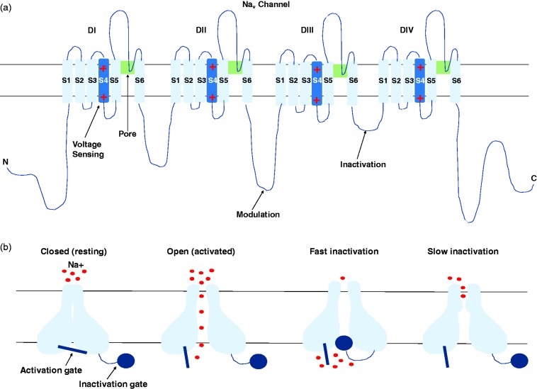
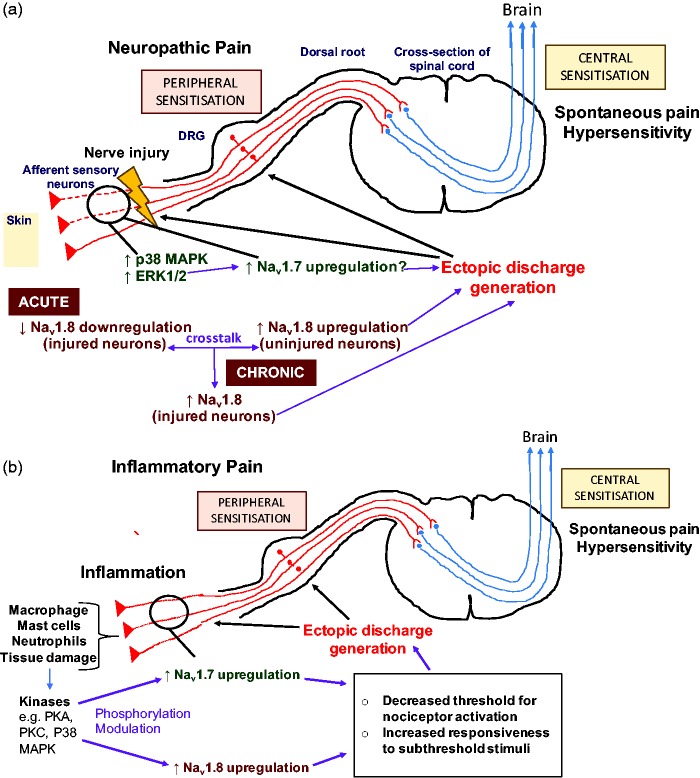
Chronic pain is a significant unmet medical problem. Current research regarding sodium channel function in pathological pain is advancing with the hope that it will enable the development of isoform-specific sodium channel blockers, a promising treatment for chronic pain. Before advancements in the pharmacological field, an elucidation of the roles of Nav1.7 and Nav1.8 in the pathophysiology of pain states is required. Thus, the aim of this report is to present what is currently known about the contributions of these sodium channel subtypes in the pathophysiology of neuropathic and inflammatory pain. The electrophysiological properties and localisation of sodium channel isoforms is discussed. Research concerning the genetic links of Nav1.7 and Nav1.8 in acquired neuropathic and inflammatory pain states from the scientific literature in this field is reported. The role of Nav1.7 and Nav1.8 in the generation and maintenance of abnormal neuronal electrogenesis and hyperexcitability highlights the importance of these channels in the development of pathological pain. However, further research in this area is required to fully elucidate the roles of Nav1.7 and Nav1.8 in the pathophysiology of pain for the development of subtype-specific sodium channel blockers.
Keywords: Nav1.7; Nav1.8; dorsal root ganglion; hyperexcitability; inflammatory pain; neuropathic pain; nociceptors; sodium channel; voltage-gated sodium channels.
Figures
Similar articles
-
Unique electrophysiological property of a novel Nav1.7, Nav1.8, and Nav1.9 sodium channel blocker, ANP-230.Biochem Biophys Res Commun. 2024 Aug 20;721:150126. doi: 10.1016/j.bbrc.2024.150126. Epub 2024 May 14. Biochem Biophys Res Commun. 2024. PMID: 38776832
-
Voltage-gated sodium channel in grasshopper mice defends against bark scorpion toxin.Science. 2013 Oct 25;342(6157):441-446. doi: 10.1126/science.1236451. Science. 2013. PMID: 24159039 Free PMC article.
-
Spider venom-derived peptide induces hyperalgesia in Nav1.7 knockout mice by activating Nav1.9 channels.Nat Commun. 2020 May 8;11(1):2293. doi: 10.1038/s41467-020-16210-y. Nat Commun. 2020. PMID: 32385249 Free PMC article.
-
Nav1.8 and Chronic Pain: From Laboratory Animals to Clinical Patients.Biomolecules. 2025 May 10;15(5):694. doi: 10.3390/biom15050694. Biomolecules. 2025. PMID: 40427587 Free PMC article. Review.
-
Pain behavior in SCN9A (Nav1.7) and SCN10A (Nav1.8) mutant rodent models.Neurosci Lett. 2021 May 14;753:135844. doi: 10.1016/j.neulet.2021.135844. Epub 2021 Mar 26. Neurosci Lett. 2021. PMID: 33775738 Review.
Cited by
-
Possible Combinatorial Utilization of Phytochemicals and Extracellular Vesicles for Wound Healing and Regeneration.Int J Mol Sci. 2024 Sep 26;25(19):10353. doi: 10.3390/ijms251910353. Int J Mol Sci. 2024. PMID: 39408681 Free PMC article. Review.
-
Cyclovirobuxine D, a cardiovascular drug from traditional Chinese medicine, alleviates inflammatory and neuropathic pain mainly via inhibition of voltage-gated Cav3.2 channels.Front Pharmacol. 2022 Dec 21;13:1081697. doi: 10.3389/fphar.2022.1081697. eCollection 2022. Front Pharmacol. 2022. PMID: 36618940 Free PMC article.
-
A Phase 1, Randomized, Double-Blind, Placebo-Controlled, Crossover Study to Evaluate the Pharmacodynamic Effects of VX-150, a Highly Selective NaV1.8 Inhibitor, in Healthy Male Adults.Pain Med. 2021 Aug 6;22(8):1814-1826. doi: 10.1093/pm/pnab032. Pain Med. 2021. PMID: 33543763 Free PMC article. Clinical Trial.
-
Profiling human iPSC-derived sensory neurons for analgesic drug screening using a multi-electrode array.Cell Rep Methods. 2025 May 19;5(5):101051. doi: 10.1016/j.crmeth.2025.101051. Epub 2025 May 13. Cell Rep Methods. 2025. PMID: 40367946 Free PMC article.
-
Emerging Molecular and Synaptic Targets for the Management of Chronic Pain Caused by Systemic Lupus Erythematosus.Int J Mol Sci. 2024 Mar 22;25(7):3602. doi: 10.3390/ijms25073602. Int J Mol Sci. 2024. PMID: 38612414 Free PMC article. Review.
References
-
- Dib-Hajj SD, Cummins TR, Black JA, Waxman SG. Sodium channels in normal and pathological pain. Annu Rev Neurosci 2010; 33: 325–347. - PubMed
-
- Wall PD, Waxman SG, Basbaum AI. Ongoing activity in peripheral nerve, III. Injury discharge. Exp Neurol 1974; 45: 578–589. - PubMed
-
- Waxman SG, Cummins TR, Dib-Hajj S, Fjell J, Black JA. Sodium channels, excitability of primary sensory neurons, and the molecular basis of pain. Muscle Nerve 1999; 22: 1177–1187. - PubMed
-
- Dib-Hajj SD, Binshtok AM, Cummins TR, Jarvis MF, Samad T, Zimmermann K. Voltage-gated sodium channels in pain states: role in pathophysiology and targets for treatment. Brain Res Rev 2009; 60: 65–83. - PubMed
Publication types
MeSH terms
Substances
LinkOut - more resources
Full Text Sources
Other Literature Sources
Medical
