Review
. 2019 May;7(3):10.1128/microbiolspec.gpp3-0062-2019.
doi: 10.1128/microbiolspec.GPP3-0062-2019.

Pathogenicity Islands and Their Role in Staphylococcal Biology

Affiliations
Review

Pathogenicity Islands and Their Role in Staphylococcal Biology

Richard P Novick. Microbiol Spectr. 2019 May.

Abstract

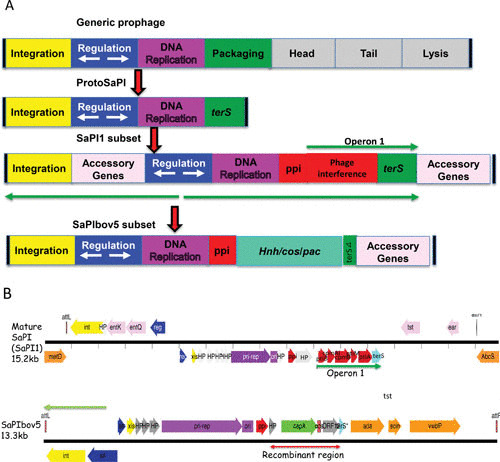
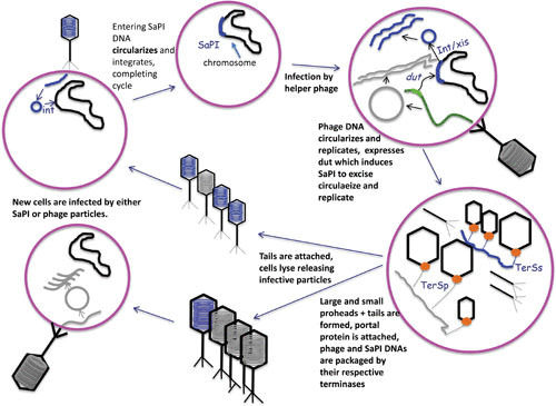
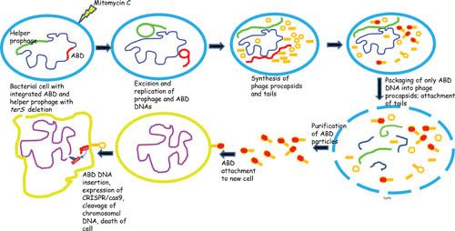
Pathogenicity islands are members of a vast collection of genomic islands that encode important virulence, antibiotic resistance and other accessory functions and have a critical role in bacterial gene transfer. Staphylococcus aureus is host to a large family of such islands, known as SaPIs, which encode super antigen and other virulence determinants, are mobilized by helper phages and transferred at extremely high frequencies. They benefit their host cells by interfering with phage predation and enhancing horizontal gene transfer. This chapter describes their life cycle, the bases of their phage interference mechanisms, their transfer system and their conversion to antibacterial agents for treatment ofstaphylococcal infections.


Figures

FIGURE 1
FIGURE 1
The SaPI genome and its putative origin. (A) Block diagrams. At the top is a typical prophage genome, and below that is the putative SaPI precursor, presumably the result of a major deletion. Next are two extant SaPI genomes. (B) Genetic maps of SaPI1 and SaPIbov5. Colors: red, interference genes; aqua, terS; orange, accessory genes; blue, regulatory genes; yellow, int/xis; gray, hypotheticals; purple, replication.
FIGURE 2
FIGURE 2
SaPI genomic pattern. At the top is the groEL region of S. aureus strain mu50, with a classical SaPI inserted at the 3′ end of groEL. The basic SaPI genes are in gray, and accessory genes at the left end are in green. Note the typical transcriptional divergence. Below is shown the corresponding region of strain JH9, in which the SaPI att site at groEL is empty.
FIGURE 3
FIGURE 3
The SaPI life cycle.
FIGURE 4
FIGURE 4
Induction, maturation, release, and cell killing by a CRISPR/cas9 ABD.
FIGURE 5
FIGURE 5
Diagrammatic representation of the ABD backbone.

References

    1. Hayes W. 1968. The Genetics of Bacteria and their Viruses, 2nd ed. Blackwell Scientific Publications, Oxford, Edinburgh, UK.
    1. Jordan E, Saedler H, Starlinger P. 1967. Strong-polar mutations in the transferase gene of the glactose operon in E coli. P. Mol Gen Genet 100:296–306. - PubMed
    1. Summers DK. 1996. The Biology of Plasmids. Blackwell Science Ltd., Oxford, UK. 10.1002/9781444313741. - DOI
    1. Blum G, Ott M, Lischewski A, Ritter A, Imrich H, Tschäpe H, Hacker J. 1994. Excision of large DNA regions termed pathogenicity islands from tRNA-specific loci in the chromosome of an Escherichia coli wild-type pathogen. Infect Immun 62:606–614. - PMC - PubMed
    1. Damle PK, Wall EA, Spilman MS, Dearborn AD, Ram G, Novick RP, Dokland T, Christie GE. 2012. The roles of SaPI1 proteins gp7 (CpmA) and gp6 (CpmB) in capsid size determination and helper phage interference. Virology 432:277–282 10.1016/j.virol.2012.05.026. [PubMed] - DOI - PMC - PubMed

Substances

LinkOut - more resources