. 2019 Aug;597(15):3817-3832.
doi: 10.1113/JP277769. Epub 2019 Jul 11.

MitoBKCa channel is functionally associated with its regulatory β1 subunit in cardiac mitochondria

Affiliations

MitoBKCa channel is functionally associated with its regulatory β1 subunit in cardiac mitochondria

Enrique Balderas et al. J Physiol. 2019 Aug.

Abstract

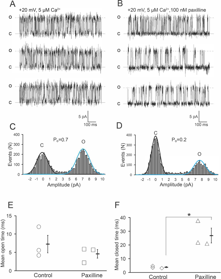
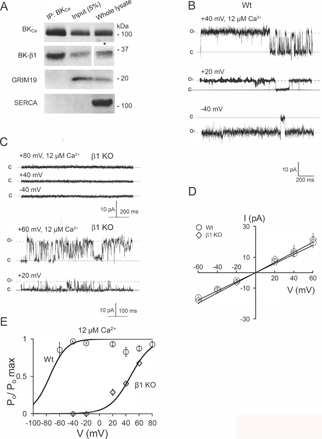
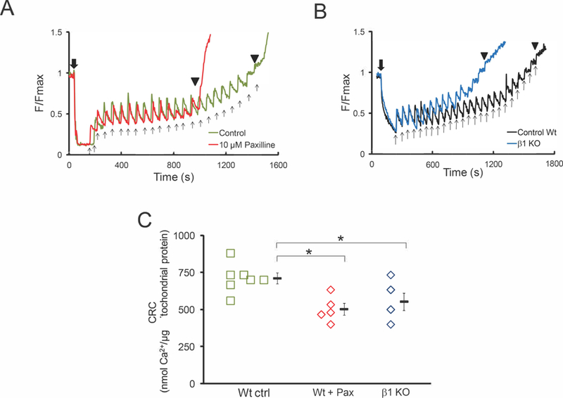
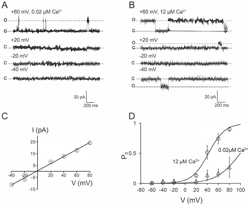
Key points: Association of plasma membrane BKCa channels with BK-β subunits shapes their biophysical properties and physiological roles; however, functional modulation of the mitochondrial BKCa channel (mitoBKCa ) by BK-β subunits is not established. MitoBKCa -α and the regulatory BK-β1 subunit associate in mouse cardiac mitochondria. A large fraction of mitoBKCa display properties similar to that of plasma membrane BKCa when associated with BK-β1 (left-shifted voltage dependence of activation, V1/2 = -55 mV, 12 µm matrix Ca2+ ). In BK-β1 knockout mice, cardiac mitoBKCa displayed a low Po and a depolarized V1/2 of activation (+47 mV at 12 µm matrix Ca2+ ) Co-expression of BKCa with the BK-β1 subunit in HeLa cells doubled the density of BKCa in mitochondria. The present study supports the view that the cardiac mitoBKCa channel is functionally modulated by the BK-β1 subunit; proper targeting and activation of mitoBKCa shapes mitochondrial Ca2+ handling.

Abstract: Association of the plasma membrane BKCa channel with auxiliary BK-β1-4 subunits profoundly affects the regulatory mechanisms and physiological processes in which this channel participates. However, functional association of mitochondrial BK (mitoBKCa ) with regulatory subunits is unknown. We report that mitoBKCa functionally associates with its regulatory subunit BK-β1 in adult rodent cardiomyocytes. Cardiac mitoBKCa is a calcium- and voltage-activated channel that is sensitive to paxilline with a large conductance for K+ of 300 pS. Additionally, mitoBKCa displays a high open probability (Po ) and voltage half-activation (V1/2 = -55 mV, n = 7) resembling that of plasma membrane BKCa when associated with its regulatory BK-β1 subunit. Immunochemistry assays demonstrated an interaction between mitochondrial BKCa -α and its BK-β1 subunit. Mitochondria from the BK-β1 knockout (KO) mice showed sparse mitoBKCa currents (five patches with mitoBKCa activity out of 28 total patches from n = 5 different hearts), displaying a depolarized V1/2 of activation (+47 mV in 12 µm matrix Ca2+ ). The reduced activity of mitoBKCa was accompanied by a high expression of BKCa transcript in the BK-β1 KO, suggesting a lower abundance of mitoBKCa channels in this genotype. Accordingly, BK-β1subunit increased the localization of BKDEC (i.e. the splice variant of BKCa that specifically targets mitochondria) into mitochondria by two-fold. Importantly, both paxilline-treated and BK-β1 KO mitochondria displayed a more rapid Ca2+ overload, featuring an early opening of the mitochondrial transition pore. We provide strong evidence that mitoBKCa associates with its regulatory BK-β1 subunit in cardiac mitochondria, ensuring proper targeting and activation of the mitoBKCa channel that helps to maintain mitochondrial Ca2+ homeostasis.

Keywords: Paxilline; cardiomyocytes; mitochondria; mitochondrial calcium retention capacity; potassium channel.


Conflict of interest statement

Competing interests

The authors declare no competing financial interests.

Figures

Figure 1
Figure 1. Preparation of mitoplast from left ventricle cardiomyocytes
A) Healthy rat cardiomyocytes isolated with a Langendorff perfusion apparatus. B) Electron microscopy image of mitoplasts (inner mitochondrial membrane devoid of outer membrane), red arrow. A low percentage of intact swollen mitochondria remained (top-left panel B). C) Mitoplasts labeled with Mitotracker Red visualized with differential interference contrast (DIC) Nomarski microscopy through a 60X objective. The arrow in C indicate a selected mitoplast for patch-clamp. D) DIC image of panel C show a borosilicate patch pipette next to a mitoplast. E) Schematic representation of the preparation of mitoplasts. In an excised patch (inside-out configuration), the bath solution represents the mitochondrial matrix.
Figure 2
Figure 2. Voltage-dependence of MitoBKCa channel.
In an excised inside-out patch, the bath solution represents the matrix side of the mitoplast. Channel activity was recorded in symmetric 150 KMeSO4. A) Single channel currents from a patch that contains at least three channels obtained at 12 µM matrix Ca2+ (c=closed state; o=open state). B) The same patch as in A exposed to 5 µM matrix Ca2+. Recordings in B show the activity of only one channel at positive membrane potentials. C) Same patch as in A and B exposed to 0.02 µM matrix Ca2+, single channel current was observed only at +80 mV. D) Plot of normalized NPo vs V. Data points were fit to a Boltzmann distribution (continuous lines) with a V1/2=−55, +42 and +70 mV at 12 (n=8), 5 (n=4), and 0.02 µM Ca2+ (n=3), respectively. Z values of 2.2, 1.4 and 1.6 were calculated for 12, 5 and 0.02 µM matrix Ca2+, respectively. E) Plots of I vs. V of mitoBKCa channels with a slope conductance of 309±19 pS (n=11). Bars represent mean and SEM here and throughout.
Figure 3
Figure 3. Paxilline reduces the open probability of mitoBKCa channels.
Single-channel currents of mitoBKCa recorded from the same mitoplast in absence (A) and in presence of paxilline (B) at the indicated matrix Ca2+ and membrane potential. Paxilline decreased the Po of the 7 pA channel from 0.77±0.1 to 0.41±0.1 (n=3) as indicated in the current amplitude histograms (C and D). Mean open time (E) and mean closed time (F) observed in 3 different patches. Paxilline augmented significantly the mean closed time of the channel (*p<0.05 Student’s t-test, n=3 hearts).
Figure 4
Figure 4. MitoBKCa alpha and auxiliary β1 subunit association in cardiac mitochondria.
A) Immunoprecipitation (IP) of BKCa alpha and BK-β1 subunit with an antibody against BKCa alpha (see methods). Western blots revealed the presence of GRIM 19 and SERCA as markers of mitochondrial and sarcoplasmic reticulum membranes (4A, lower panels). *The signal of BK-β1 in the whole lysate was detected with chemiluminescent (ECL) substrate Supersignal™ West Femto Maximun sensitivity (see methods). Taken together, these results indicates an association between mitoBKCa and its auxiliary β1 subunit. Having demonstrated this, we characterized the biophysical properties of mitoBKCa in Wt and β1-KO cardiac mitoplasts. B) Single-channel currents of mitoBKCa recorded from Wt mitoplasts. The Po of the channel was 0.9 ± 0.08, n=4, in average at +80 mV and the indicated matrix Ca2+. C) Representative traces of a mitoplast from the β1 KO, were no single-channel current was detected at the indicated voltages and matrix Ca2+ (upper traces). Few mitoplasts (5 out of 28, mitoplasts, n=5 hearts) showed mitoBKCa channel activity with a Po of 0.4 ± 0.004, n=3 at +40 mV in the indicated matrix Ca2+. D) Plots of I vs. V at 12 µM matrix Ca2+ from Wt and β1 KO mitoplasts (data from 6 and 3 patches, respectively). A slope conductance of 302±26 (n=4) and 335 ±15 pS (n=3) was calculated for Wt and β1 KO, respectively. E) Plots of Po vs. V for mitoBKCa channels, data points represent the average of 4 and 3 mitoplasts for Wt and BK-β1 KO, respectively. Bars SEM.
Figure 5
Figure 5. Paxilline reversibly blocks mouse cardiac mitoBKCa channel
Single channel current in absence (A) and in presence (B) of paxilline at the indicated voltage and matrix Ca2+. C) Current amplitude histogram of the 6 pA channel recorded in A, paxilline reduced the Po from 0.7±0.1 to 0.2±0.09 (n=3) (D). E) Single channel current recorded after the patch was perfused again with 12 µM matrix Ca2+. F) Current amplitude histogram that shows the reappearance of the 6 pA current (Po=0.4) after washing out paxilline.
Figure 6
Figure 6. Localization of BKDEC and β1 subunit in HeLa cells.
A) Cultured cells co-transfected with BKDEC alpha+β1subunit, loaded with mitotracker red, and DAPI B) β1-flag labeled with an anti-flag C) BKDEC labeled with anti-HA. D) Merge of mitotracker red, BKDEC and β1. E) Cross-correlation index (CCi), relative to mitotracker red signal, calculated from cells overexpressing BKDEC alone (Cci=0.53±0.05) and co-expressed with β1 subunit (Cci=0.77±0.01, n=11 cells from 3 different preparations).*p<0.05.
Figure 7
Figure 7. Mitochondrial calcium uptake is impaired by Paxilline and by genetic ablation of β1-subunit
A) Paxilline reduces the amount of mitochondrial Ca2+ necessary to induce formation and opening of mPTP. B) Mitochondria from the β1 KO required less Ca2+ than mitochondria from the Wt to trigger the opening of mPTP. C) Calcium retention capacity values obtained in A for paxilline and in B for the β1-KO mitochondria. Bars SEM, (*p<0.05 Student’s t-test). Black downward arrow, addition of mitochondria. Black upward arrows, 10 nmol CaCl2 pulses. (▼) opening of mPTP.
Figure 8
Figure 8. A different population of mitoBKCa in cardiac mitochondria
A) Single channel currents recorded in 0.02 µM matrix Ca2+ at the indicated voltages. B) Same patch as in A exposed to 12 µM matrix Ca2+. C) Plot of I vs. V, a slope conductance of 284 ± 6 pS (n=7) was calculated. D) Plots of Po vs. V from the currents observed in A and B, data points represent an average of ≥4 different patches at the indicated matrix Ca2+.

References

    1. Aon MA, Cortassa S, Wei AC, Grunnet M & O’Rourke B. (2010). Energetic performance is improved by specific activation of K+ fluxes through K(Ca) channels in heart mitochondria. Biochim Biophys Acta 1797, 71–80. - PMC - PubMed
    1. Balderas E, Zhang J, Stefani E & Toro L. (2015). Mitochondrial BKCa channel. Front Physiol 6, 104. - PMC - PubMed
    1. Bao L & Cox DH. (2005). Gating and ionic currents reveal how the BKCa channel’s Ca2+ sensitivity is enhanced by its beta1 subunit. J Gen Physiol 126, 393–412. - PMC - PubMed
    1. Barrett JN, Barrett EF & Dribin LB. (1981). Calcium-dependent slow potassium conductance in rat skeletal myotubes In press 82. - PubMed
    1. Bautista L, Castro MJ, Lopez-Barneo J & Castellano A. (2009). Hypoxia inducible factor-2alpha stabilization and maxi-K+ channel beta1-subunit gene repression by hypoxia in cardiac myocytes: role in preconditioning. Circ Res 104, 1364–1372. - PubMed

Publication types

MeSH terms

Substances