Review
. 2019 Jun 6;74(5):866-876.
doi: 10.1016/j.molcel.2019.04.027.

Replication-Coupled DNA Repair

Affiliations
Review

Replication-Coupled DNA Repair

David Cortez. Mol Cell. .

Abstract

The replisome quickly and accurately copies billions of DNA bases each cell division cycle. However, it can make errors, especially when the template DNA is damaged. In these cases, replication-coupled repair mechanisms remove the mistake or repair the template lesions to ensure high fidelity and complete copying of the genome. Failures in these genome maintenance activities generate mutations, rearrangements, and chromosome segregation problems that cause many human diseases. In this review, I provide a broad overview of replication-coupled repair pathways, explaining how they fix polymerase mistakes, respond to template damage that acts as obstacles to the replisome, deal with broken forks, and impact human health and disease.

Keywords: DNA damage; DNA replication; break-induced replication; cancer; crosslink repair; excision repair; fork protection; fork reversal; mismatch repair; replication stress.


Conflict of interest statement

Declaration of Interests

The authors declare no competing interests. eTOC blurb: Replication-coupled DNA repair minimizes replication errors and prevents disease. This review discusses these mechanisms and provides perspective on important concepts and outstanding questions.

Figures
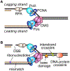
Fig. 1.
Fig. 1.
DNA lesions that are resolved by replication-coupled DNA repair pathways. (A) A simplified diagram of a eukaryotic replication fork. (B) Lesions generated or encountered by the replisome.
Fig. 2.
Fig. 2.
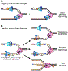
Differences in responses to leading and lagging strand base damage. (A) Most lagging strand damage will generate ssDNA gaps but may not stall DNA elongation due to rapid repriming by Pola-primase. (B) Translesion synthesis, PrimPol-dependent repriming, and fork reversal are three pathways to overcome leading strand lesions that cause uncoupling of replisome activities.
Fig. 3.
Fig. 3.
Illustration of the complexities of fork reversal. Fork reversal places a template DNA lesion back into duplex DNA where it can be removed by excision repair. The ATP-dependent DNA translocases, SMARCAL1, HLTF, and ZRANB3 are regulated by RPA, 3’ DNA ends, and poly-ubiquitylated PCNA respectively to catalyze fork reversal. In some cases these enzymes may work sequentially, although the order of action depicted in the diagram is speculative. ATR signaling controls SMARCAL1 and also may stimulate reversal of undamaged forks. RAD51 acts through an unknown mechanism to promote reversal, and then RAD51 filaments stabilized by BRCA2 and additional homology directed repair (HDR) proteins prevent nucleases from degrading the nascent DNA strands.
Fig. 4.
Fig. 4.
Possible replication-coupled ICL repair mechanisms. (A) Two converging forks approach the ICL. Short-chain TRAIP-dependent ubiquitylation promotes NEIL3 recruitment and strand unhooking. Long-chain ubiquitylation causes replisome unloading followed by fork reversal and unhooking by endonuclease incisions via the Fanconi anaemia pathway. (B) ICLs can be traversed with the help of the accessory DNA helicases FANCM and BLM. Remodeling of the CMG helicase may allow it to move past the ICL. Repriming then allows continued DNA synthesis leaving the ICL to be repaired through the Fanconi anaemia pathway.
Fig. 5.
Fig. 5.
Four mechanisms for generating a single-ended double-strand break at replications forks. Single-ended breaks are repaired by break induced replication mechanisms that depend on an alternative replisome.

References

    1. Amunugama R, Willcox S, Wu RA, Abdullah UB, El-Sagheer AH, Brown T, McHugh PJ, Griffith JD, and Walter JC (2018). Replication Fork Reversal during DNA Interstrand Crosslink Repair Requires CMG Unloading. Cell Rep 23, 3419–3428. - PMC - PubMed
    1. Anand RP, Lovett ST, and Haber JE (2013). Break-induced DNA replication. Cold Spring Harbor perspectives in biology 5, a010397. - PMC - PubMed
    1. Badu-Nkansah A, Mason AC, Eichman BF, and Cortez D (2016). Identification of a Substrate Recognition Domain in the Replication Stress Response Protein Zinc Finger Ran- binding Domain-containing Protein 3 (ZRANB3). J Biol Chem 291, 8251–8257. - PMC - PubMed
    1. Bansbach CE, Betous R, Lovejoy CA, Glick GG, and Cortez D (2009). The annealing helicase SMARCAL1 maintains genome integrity at stalled replication forks. Genes Dev 23, 2405–2414. - PMC - PubMed
    1. Bass TE, and Cortez D (2019). Quantitative phosphoproteomics reveals mitotic function of the ATR activator ETAA1. J Cell Biol 218, 1235–1249. - PMC - PubMed

Publication types