. 2020 Aug 1;12(8):654-665.
doi: 10.1093/jmcb/mjz051.

Mitotic motor CENP-E cooperates with PRC1 in temporal control of central spindle assembly

Affiliations

Mitotic motor CENP-E cooperates with PRC1 in temporal control of central spindle assembly

Xu Liu et al. J Mol Cell Biol. .

Abstract

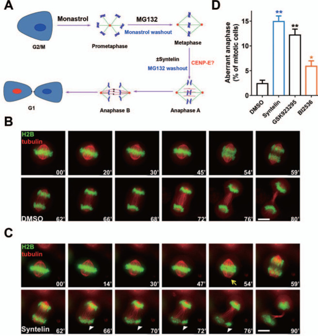
Error-free cell division depends on the accurate assembly of the spindle midzone from dynamic spindle microtubules to ensure chromatid segregation during metaphase-anaphase transition. However, the mechanism underlying the key transition from the mitotic spindle to central spindle before anaphase onset remains elusive. Given the prevalence of chromosome instability phenotype in gastric tumorigenesis, we developed a strategy to model context-dependent cell division using a combination of light sheet microscope and 3D gastric organoids. Light sheet microscopic image analyses of 3D organoids showed that CENP-E inhibited cells undergoing aberrant metaphase-anaphase transition and exhibiting chromosome segregation errors during mitosis. High-resolution real-time imaging analyses of 2D cell culture revealed that CENP-E inhibited cells undergoing central spindle splitting and chromosome instability phenotype. Using biotinylated syntelin as an affinity matrix, we found that CENP-E forms a complex with PRC1 in mitotic cells. Chemical inhibition of CENP-E in metaphase by syntelin prevented accurate central spindle assembly by perturbing temporal assembly of PRC1 to the midzone. Thus, CENP-E-mediated PRC1 assembly to the central spindle constitutes a temporal switch to organize dynamic kinetochore microtubules into stable midzone arrays. These findings reveal a previously uncharacterized role of CENP-E in temporal control of central spindle assembly. Since CENP-E is absent from yeast, we reasoned that metazoans evolved an elaborate central spindle organization machinery to ensure accurate sister chromatid segregation during anaphase and cytokinesis.

Keywords: CENP-E; PRC1; cell division; central spindle; organoids; syntelin.


Figures

Figure 1
Figure 1
Modeling mitosis using a light sheet microscopy and chemical probes. (A) Cartoon presentation of generation of patient-derived gastric organoids. The organoid model provides a unique platform to delineate cellular dynamics during cell division in native tissue and imply synthetic lethal strategy to identify individualized medication. (B) 3D projections of gastric organoids montages from four different focal planes (Z = 92, 126, 142, and 246, respectively) that were labeled with tubulin and DAPI. The magnified images provide apparent configuration of chromosome relative to mitotic spindle. The lower imaging toxicity combined with unlimited sample depth has established light sheet microscope as a unique platform for studying molecular and cellular dynamics in live tissue. Aberrant chromosome movements such as misalignments are indicated by arrows. Scale bar, 100 μm in organoids imaging; 10 μm in magnified montage of individual mitotic cells. (C) Statistical analyses of mitotic index from different chemical probes such as CENP-E inhibitors. Note that CENP-E inhibition by syntelin and GSK923295 exhibits chronic mitotic arrest. Data represent mean ± SEM from three independent experiments. Statistical significance was determined by two-sided t-test (**P < 0.01; n = 21 for each group). (D) Magnified montage of an anaphase cells treated with syntelin. The magnified anaphase cell exhibits typical lagging chromosome (arrow). (E) Statistical analyses of aberrant anaphase cells from organoids treated with mitotic modulators such as CENP-E inhibitors. Note that CENP-E inhibition by syntelin and GSK923295 significantly increased the number of cells exhibiting aberrant anaphase chromosomes (**P < 0.01; n = 23 for each group). Data represent mean ± SEM from three independent experiments.
Figure 2
Figure 2
CENP-E kinesin activity is essential for accurate anaphase onset. (A) Schematic presentation of delineating temporal function of CENP-E in mitosis. HeLa cells were synchronized at metaphase using chemical inhibitors and exposed to syntelin treatment. (B) Representative montage from live HeLa cells expressing GFP-H2B and mCherry-tubulin. HeLa cells were transfected to express GFP-H2B and mCherry-tubulin for 24 h prior to MG132 synchronization and real-time imaging experimentation as illustrated in A. Note that the cell was treated with DMSO. Scale bar, 10 μm. (C) Representative montage from live HeLa cells expressing GFP-H2B and mCherry-tubulin with syntelin treatment. HeLa cells were transfected to express GFP-H2B and mCherry-tubulin for 24 h prior to MG132 synchronization and parallel experimentation with syntelin as in B. Note that the cell treated with 1 μM syntelin at the metaphase exhibited aberrant metaphase–anaphase transition with chromatid split phenotype. Note that arrowheads indicate the chromatid split phenotype. Scale bar, 10 μm. (D) Quantification of aberrant anaphase phenotypes of live HeLa cells treated with DMSO, syntelin (1 μM), GSK923295 (100 nM), or BI2536 (100 nM). Data represent mean ± SEM from three independent experiments. Statistical significance was tested by two-sided t-test (*P < 0.05; **P < 0.01; n = 25 for each group).
Figure 3
Figure 3
CENP-E kinesin is required to organize PRC1 to the spindle midzone. (A) Representative montage from live HeLa cells expressing dual-color reporters (GFP-PRC1 and mCherry-H2B). HeLa cells were transfected to express GFP-PRC1 and mCherry-H2B for 24 h prior to synchronization at the metaphase for the real-time experimentation. Note that the cell treated with DMSO progressed into anaphase with accurate chromatid segregation. (B) Representative montage from live HeLa cells expressing dual-color reporters (GFP-PRC1 and mCherry-H2B) with syntelin treatment. HeLa cells were transfected to express GFP-PRC1 and mCherry-H2B for 24 h prior to synchronization at the metaphase for syntelin treatment and the real-time experimentation. Note that the cell treated with 1 μM syntelin at the metaphase exhibited perturbation in establishment of central spindle midzone and errors in sister chromatid separation and segregation. (C) Representative montage from live HeLa cells expressing GFP-PRC1. Note that PRC1-labeled central spindle was organized and dynamically concentrated into the midbody in DMSO-treated cells. (D) Representative montage from live HeLa cells expressing GFP-PRC1. Note that the cell was treated with 1 μM syntelin at the metaphase and syntelin treatment inhibited the formation of PRC1-organized anti-parallel central spindle. (E) Quantification of mitotic phenotypes of live HeLa cells treated with DMSO, syntelin (1 μM), GSK923295 (100 nM), or reversine (300 nM). Data represent mean ± SEM from three independent experiments. Statistical significance was tested by two-sided t-test (**P < 0.01; n = 50).
Figure 4
Figure 4
CENP-E kinesin is essential for accurate establishment of central spindle. (A) HeLa cells treated with DMSO and syntelin (1 μM) were subjected to immunofluorescence assay for CENP-E (red), tubulin (green), and DAPI. Note that syntelin-treated cells exhibit aberrant central spindle revealed by tubulin staining (arrow). Scale bar, 10 μm. (B) HeLa cells treated with DMSO and syntelin (1 μM) were subjected to immunofluorescence assay for MKLP1 (red), tubulin (green), and DAPI. Note that syntelin-treated cells exhibit virtually no MKLP1 (arrow). Scale bar, 10 μm. (C) Quantification of central spindle location of CENP-E, MKLP1, and PRC1 in HeLa cells treated with DMSO, syntelin (1 μM), GSK923296 (100 nM), and reversine (300 nM), respectively. Data represent mean ± SEM from three independent experiments. Statistical significance was tested by two-sided t-test (**P < 0.01; n = 50). (D) Inhibition of CENP-E perturbed central spindle organization. HeLa cells were processed as described in Materials and methods prior to electron microscopic analyses of anaphase central spindle of HeLa cells treated with DMSO and syntelin (1 μM). (a) Lower magnification of view of a late anaphase HeLa cell bearing elongated spindle poles, labeled with asterisks. Interzonal microtubules are readily seen (boxed). Scale bar, 2 μm. (b) Magnified view of the boxed region in a, showing that anti-parallel microtubules are readily apparent (red arrowhead). Arrows indicate CENP-E gold particle deposition. Scale bar, 200 nm. (c) Lower magnification of view of a late anaphase HeLa cell treated with syntelin, labeled with asterisks. Interzonal microtubules are perturbed without apparent central spindle (boxed). Scale bar, 2 μm. (d) Magnified view of the boxed region in c, showing that anti-parallel microtubules are collapsed (red arrow). Scale bar, 200 nm. Note that syntelin treatment prevents the establishment of anti-parallel microtubules that fail to power the elongation of central spindles. (E) Schematic presentation of working model to account for CENP-E function in central spindle anti-parallel microtubule establishment and organization. We proposed that CENP-E organizes anti-parallel microtubules and powers microtubule sliding in overlapped midzone.
Figure 5
Figure 5
CENP-E forms a functional complex with PRC1 in mitosis. (A) Schematic drawing of syntelin-affinity matrix-based discovery of CENP-E binders. In brief, clarified mitotic HeLa cell lysates were subjected to binding to biotinylated syntelin followed by absorption with avidin-sepharose beads. The bound materials after extensive wash will be resolved by SDS-PAGE followed by mass spectrometric identification. (B) Representative SDS-PAGE analyses from syntelin-affinity purification. The clarified mitotic HeLa cell lysates were used as inputs for incubation of biotinylated syntelin followed by isolation with avidin-based affinity matrix. The proteins bound to avidin matrix were eluted with syntelin and resolved by SDS-PAGE followed by Coomassie Brilliant Blue staining. Mass spectrometric analyses indicate that CENP-E and PRC1 were recovered from avidin matrix. (C) Representative western blotting analyses for proteins isolated by syntelin-affinity purification. Note that BubR1, CENP-E, and PRC1 were isolated by syntelin–biotin–avidin matrix. (D) Identification of PRC1 as a cognate binding protein of CENP-E. The clarified mitotic HeLa cell lysates were used as inputs for incubation of anti-CENP-E antibodies followed by Protein A/G affinity chromatography. Immunoprecipitation experiment showed that endogenous PRC1 binds to CENP-E. (E) Working model accounts for novel CENP-E function in facilitating an initial central spindle establishment via a temporally controlled CENP-E–PRC1 interaction. CENP-E is situated on the outermost surface of the kinetochore during initial lateral microtubule attachment. After biorientation, CENP-E links kinetochores to spindle microtubules to achieve metaphase alignment. At metaphase, CENP-E releases and redistributes to midzone with PRC1 where they organize interzonal microtubules and power microtubule sliding that leads to pole-to-pole separation. It remains elusive as how the interaction of CENP-E with PRC1 guides the central spindle elongation and resolution in telophase.

References

    1. Adams, G., Zhou, J., Wang, W., et al. (2016). Microtubule plus-end tracking protein TIP150 interacts with cortactin to steer directional cell migration. J. Biol. Chem. 291, 20692–20706. - PMC - PubMed
    1. Akhmanova, A., and Steinmetz, M.O. (2008). Tracking the ends: a dynamic protein network controls the fate of microtubule tips. Nat. Rev. Mol. Cell Biol. 9, 309–322. - PubMed
    1. Akram, S., Yang, F., Li, J., et al. (2018). LRIF1 interacts with HP1alpha to coordinate accurate chromosome segregation during mitosis. J. Mol. Cell Biol. 10, 527–538. - PMC - PubMed
    1. Bakhoum, S.F., Ngo, B., Laughney, A.M., et al. (2018). Chromosomal instability drives metastasis through a cytosolic DNA response. Nature 553, 467–472. - PMC - PubMed
    1. Bao, X., Liu, H., Liu, X., et al. (2018). Mitosis-specific acetylation tunes Ran effector binding for chromosome segregation. J. Mol. Cell Biol. 10, 18–32. - PMC - PubMed

Publication types