Review
. 2019 Jun 6;20(11):2776.
doi: 10.3390/ijms20112776.

A Critical Review of Electroporation as A Plasmid Delivery System in Mouse Skeletal Muscle

Affiliations
Review

A Critical Review of Electroporation as A Plasmid Delivery System in Mouse Skeletal Muscle

Emilia Sokołowska et al. Int J Mol Sci. .

Abstract

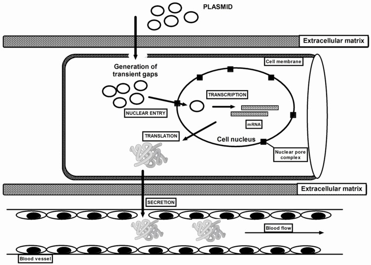
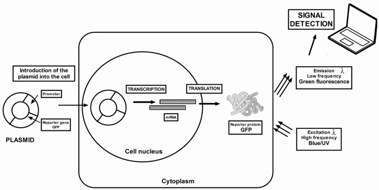
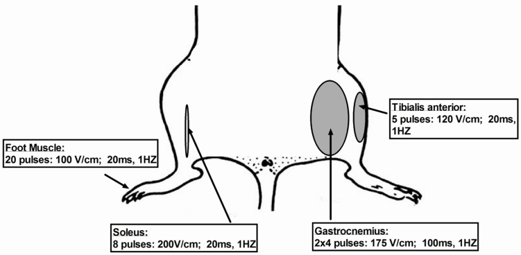
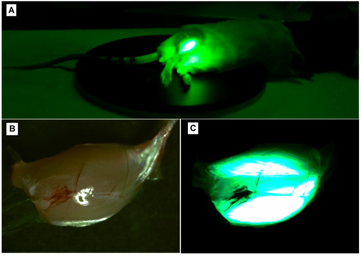
The gene delivery to skeletal muscles is a promising strategy for the treatment of both muscular disorders (by silencing or overexpression of specific gene) and systemic secretion of therapeutic proteins. The use of a physical method like electroporation with plate or needle electrodes facilitates long-lasting gene silencing in situ. It has been reported that electroporation enhances the expression of the naked DNA gene in the skeletal muscle up to 100 times and decreases the changeability of the intramuscular expression. Coelectransfer of reporter genes such as green fluorescent protein (GFP), luciferase or beta-galactosidase allows the observation of correctly performed silencing in the muscles. Appropriate selection of plasmid injection volume and concentration, as well as electrotransfer parameters, such as the voltage, the length and the number of electrical pulses do not cause long-term damage to myocytes. In this review, we summarized the electroporation methodology as well as the procedure of electrotransfer to the gastrocnemius, tibialis, soleus and foot muscles and compare their advantages and disadvantages.

Keywords: animal models; electroporation; gene electrotransfer; mouse; muscle; non-viral; plasmids; silencing.


Conflict of interest statement

The authors declare no conflict of interest.

Figures

Figure 1
Figure 1
Schematic diagram of proposed plasmid DNA delivery with the use of electroporation. Plasmid DNA (pDNA) must overcome barriers, including extracellular matrix, cell membrane, cytoplasm and nuclear membrane before reaching the nucleus. However, applying an electric current with an appropriate voltage facilitate the transport of pDNA through biological membranes. Plasmids penetrate into the target cell cytoplasm through the transiently formed gaps in the sarcolemma. DNA in the cytoplasm is exposed to degradation by cytoplasmic nucleases. Finally, the DNA through nuclear pore complexes has to penetrate the nuclear membrane to enter the nucleus where the transcription occurs. The delivered plasmid DNA may silence genes or be transcribed into the missing protein and then secreted into the bloodstream.
Figure 2
Figure 2
Diagram of the bioluminescence imaging technique using the green fluorescent protein (GFP) reporter gene. The GFP reporter gene is directly attached to the gene of interest to produce gene fusions. The two genes are located under the same promoter elements and are transcribed into a single messenger RNA molecule. The mRNA is then translated into a protein. The protein-GFP hybrid transcribed from the reporter construct possesses a protein linked to GFP. Based on the fluorescence, it can be concluded that the protein is everywhere where green fluorescence occurs.
Figure 3
Figure 3
Schematic representation of electroporation parameters.
Figure 4
Figure 4
The photographs made by our research team show the bioluminescence of GFP as a reporter protein 3 days after electroporation (A) of tibialis muscle in vivo in a living mouse and (C) ex vivo in gastrocnemius muscle. The green fluorescence of muscle fibres can be observed through the skin on the same animal by means of time-lapse fluorescence imaging (A). GFP emits green light following excitation by wavelength in the UV range. (B) shows muscle viewed in the range of visible light.

References

    1. Gothelf A., Gehl J. Gene electrotransfer to skin; review of existing literature and clinical perspectives. Curr. Gene Ther. 2010;10:287–299. doi: 10.2174/156652310791823443. - DOI - PubMed
    1. Golzio M., Rols M.P., Teissie J. In vitro and in vivo electric field-mediated permeabilization, gene transfer and expression. Methods. 2004;32:126–135. doi: 10.1016/j.ymeth.2003.11.003. - DOI - PubMed
    1. Abdulhaqq S.A., Weiner D.B. DNA vaccines: Developing new strategies to enhance immune responses. Immunol. Res. 2008;42:219–232. doi: 10.1007/s12026-008-8076-3. - DOI - PubMed
    1. Ragot T., Vincent N., Chafey P., Vigne E., Gilgenkrantz H., Couton D., Cartaud J., Briand P., Kaplan J.C., Perricaudet M. Efficient adenovirus-mediated transfer of a human minidystrophin gene to skeletal muscle of mdx mice. Nature. 1993;361:647–650. doi: 10.1038/361647a0. - DOI - PubMed
    1. Levy M.Y., Barron L.G., Meyer K.B., Szoka F.C., Jr. Characterization of plasmid DNA transfer into mouse skeletal muscle: Evaluation of uptake mechanism, expression and secretion of gene products into blood. Gene Ther. 1996;3:201–211. - PubMed

MeSH terms

Substances

LinkOut - more resources