. 2019 Jun 6;24(11):2138.
doi: 10.3390/molecules24112138.

Epigallocatechin-3-Gallate Prevents Acute Gout by Suppressing NLRP3 Inflammasome Activation and Mitochondrial DNA Synthesis

Affiliations

Epigallocatechin-3-Gallate Prevents Acute Gout by Suppressing NLRP3 Inflammasome Activation and Mitochondrial DNA Synthesis

Hye Eun Lee et al. Molecules. .

Abstract

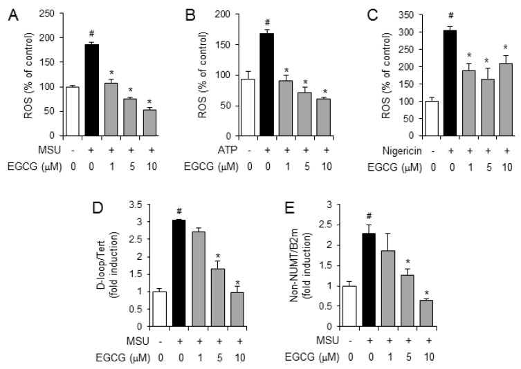
Gout is a chronic inflammatory disease evoked by the deposition of monosodium urate (MSU) crystals in joint tissues. The nucleotide-binding oligomerization domain (NOD)-like receptor (NLR) family pyrin domain containing 3 (NLRP3) inflammasome is responsible for the gout inflammatory symptoms induced by MSU crystals. We investigated whether epigallocatechin-3-gallate (EGCG) suppresses the activation of the NLRP3 inflammasome, thereby effectively preventing gouty inflammation. EGCG blocked MSU crystal-induced production of caspase-1(p10) and interleukin-1β in primary mouse macrophages, indicating its suppressive effect on the NLRP3 inflammasome. In an acute gout mouse model, oral administration of EGCG to mice effectively alleviated gout inflammatory symptoms in mouse foot tissue injected with MSU crystals. The in vivo suppressive effects of EGCG correlated well with the suppression of the NLRP3 inflammasome in mouse foot tissue. EGCG inhibited the de novo synthesis of mitochondrial DNA as well as the production of reactive oxygen species in primary mouse macrophages, contributing to the suppression of the NLRP3 inflammasome. These results show that EGCG suppresses the activation of the NLRP3 inflammasome in macrophages via the blockade of mitochondrial DNA synthesis, contributing to the prevention of gouty inflammation. The inhibitory effects of EGCG on the NLRP3 inflammasome make EGCG a promising therapeutic option for NLRP3-dependent diseases such as gout.

Keywords: gout; green tea; inflammasome; innate immunity; macrophages; mitochondria; reactive oxygen species.


Conflict of interest statement

The authors declare no conflict of interest.

Figures

Figure 1
Figure 1
Epigallocatechin-3-gallate (EGCG) suppresses the activation of the (NOD)-like receptor (NLR) family pyrin domain containing 3 (NLRP3) inflammasome in primary macrophages. Lipopolysaccharide (LPS)-primed bone marrow-derived primary macrophages were treated with EGCG for 1 h and then stimulated with monosodium uric acid (MSU) crystals (500 μg/mL for 6 h), adenosine triphosphate (ATP) (5 mM for 1 h), or nigericin (10 μM; 2 h for E and 16 h for F) in the presence or absence of EGCG as indicated. (A,C,E) Cell culture supernatants (SUP) and cell lysates (LYS) were immunoblotted for pro-caspase-1, caspase-1(p10), pro-IL-1β, and IL-1β. In the bar graph, the band density of caspase-1(p10) and cleaved IL-1β normalized to corresponding pro-caspase-1 and pro-IL-1β, was expressed as means ± SD (n = 3). #, significantly different from vehicle group, p < 0.05; *, significantly different from MSU, ATP, or nigericin alone, p < 0.05. (B,D,F) Cell culture supernatants were analyzed for secreted IL-1β by ELISA. The values represent the means ± SD (n = 3). #, significantly different from vehicle group, p < 0.05; *, significantly different from MSU, ATP, or nigericin alone, p < 0.05. N.D., not detected. IL: interleukin.
Figure 2
Figure 2
Oral administration of EGCG suppresses acute gout symptoms induced by MSU crystal injection in mice. Mice were orally administered EGCG (1, 10, and 30 mg/kg) or vehicle (0.02% dimethyl sulfoxide (DMSO) in water). After 1 h, MSU crystals (2 mg/0.1 mL of phosphate-buffered saline (PBS)/mouse) or PBS was subcutaneously injected into the footpad of the right hindfoot of each mouse. After 24 h, the footpad tissues were collected for further analysis. (A) Time course of footpad thickness gain compared with the footpad thickness at the 0 h time point per group. (B) Representative picture of hematoxylin and eosin staining of the hind feet. Infiltrating neutrophils in the hindfoot tissue appear as purple dots. (C) Representative pictures of immunohistochemistry staining of foot tissues with myeloperoxidase (MPO) (200×). (D) Supernatants of the foot tissue homogenates were analyzed for IL-6 by ELISA. The values in the line and bar graphs represent the means ± SD (n = 6 mice/group). #, significantly different from vehicle group, p < 0.05; *, significantly different from MSU alone, p < 0.05.
Figure 3
Figure 3
Oral administration of EGCG attenuates NLRP3 inflammasome activation in an acute gout mouse model. The foot tissue samples analyzed are the same as those used in Figure 2. (A) Supernatants from the foot tissue homogenates were analyzed for pro-caspase-1, caspase-1(p10), pro-IL-1β, and IL-1β by immunoblotting. # indicates an individual sample. (B) The foot tissue homogenates were analyzed for IL-1β by ELISA. The values represent the means ± SD (n = 6 mice/group). #, significantly different from vehicle group, p < 0.05; *, significantly different from MSU alone, p < 0.05. (C) Representative pictures of immunohistochemistry staining of foot tissues for caspase-1 and IL-1β (200×).
Figure 4
Figure 4
EGCG does not directly associate with NLRP3 and apoptosis-associated speck-like protein containing a CARD (ASC) or inhibit caspase-1 enzyme activity. (A,B) Sensorgrams were obtained by SPR analysis to determine the interaction between EGCG and recombinant (A) NLRP3-PYD or (B) ASC. Different concentrations of EGCG are presented as an overlay plot aligned at the start of injection. Table of kinetic parameters obtained from the SPR analysis and calculated by Biocore T200 evaluation software. (C) An in vitro assay for caspase-1 enzymatic activity was performed using a fluorometric caspase-1 assay kit with recombinant caspase-1 (rCaspase-1) in the presence or absence of EGCG or Z-YVAD-FMK. The values represent the means ± SD (n = 3). #, significantly different from vehicle, p < 0.05; *, significantly different from rCaspase-1 alone, p < 0.05.
Figure 5
Figure 5
EGCG suppresses the production of reactive oxygen species and the de novo synthesis of mitochondrial DNA induced by NLRP3 inflammasome activators in primary macrophages. (AC) Bone marrow-derived primary mouse macrophages were pretreated with an oxidative stress indicator, H2DCFDA (10 μM), for 1 h and further stimulated with monosodium uric acid (MSU) crystals (500 μg/mL), ATP (5 mM), or nigericin (10 μM) for 1 h in the presence or absence of EGCG. Reactive oxygen species (ROS) levels are presented as a percentage of the control. (D,E) LPS-primed bone marrow-derived primary mouse macrophages were stimulated with MSU crystals (500 μg/mL) for 6 h in the absence or presence of EGCG. The levels of mitochondrial DNA were determined by quantitative PCR. The values represent the means ± SD (n = 3); #, significantly different from vehicle group, p < 0.05; *, significantly different from MSU, ATP, or nigericin alone, p < 0.05.

References

    1. Wallace K.L., Riedel A.A., Joseph-Ridge N., Wortmann R. Increasing prevalence of gout and hyperuricemia over 10 years among older adults in a managed care population. J. Rheumatol. 2004;31:1582–1587. - PubMed
    1. Shi Y., Mucsi A.D., Ng G. Monosodium urate crystals in inflammation and immunity. Immunol. Rev. 2010;233:203–217. doi: 10.1111/j.0105-2896.2009.00851.x. - DOI - PubMed
    1. Martinon F., Petrilli V., Mayor A., Tardivel A., Tschopp J. Gout-associated uric acid crystals activate the NALP3 inflammasome. Nature. 2006;440:237–241. doi: 10.1038/nature04516. - DOI - PubMed
    1. Martinon F., Burns K., Tschopp J. The inflammasome: A molecular platform triggering activation of inflammatory caspases and processing of proIL-beta. Mol. Cell. 2002;10:417–426. doi: 10.1016/S1097-2765(02)00599-3. - DOI - PubMed
    1. Lee H.E., Yang G., Kim N.D., Jeong S., Jung Y., Choi J.Y., Park H.H., Lee J.Y. Targeting ASC in NLRP3 inflammasome by caffeic acid phenethyl ester: A novel strategy to treat acute gout. Sci. Rep. 2016;6:38622. doi: 10.1038/srep38622. - DOI - PMC - PubMed

MeSH terms