. 2019 Jun 6;11(6):523.
doi: 10.3390/v11060523.

Surveillance for Adenoviruses in Bats in Italy

Affiliations

Surveillance for Adenoviruses in Bats in Italy

Georgia Diakoudi et al. Viruses. .

Abstract

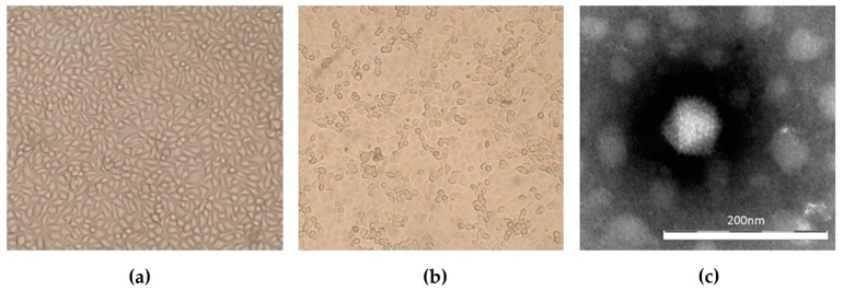
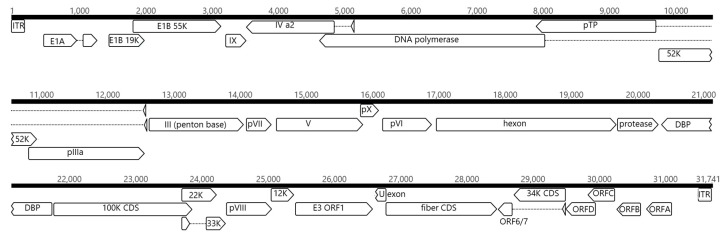
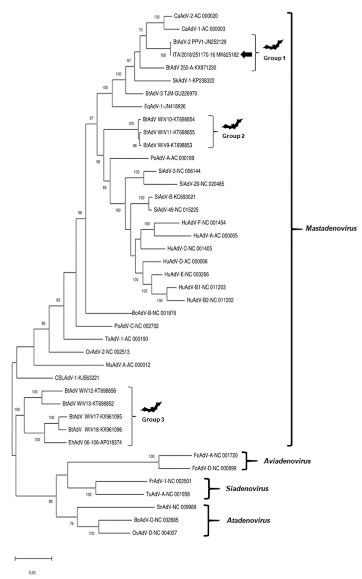
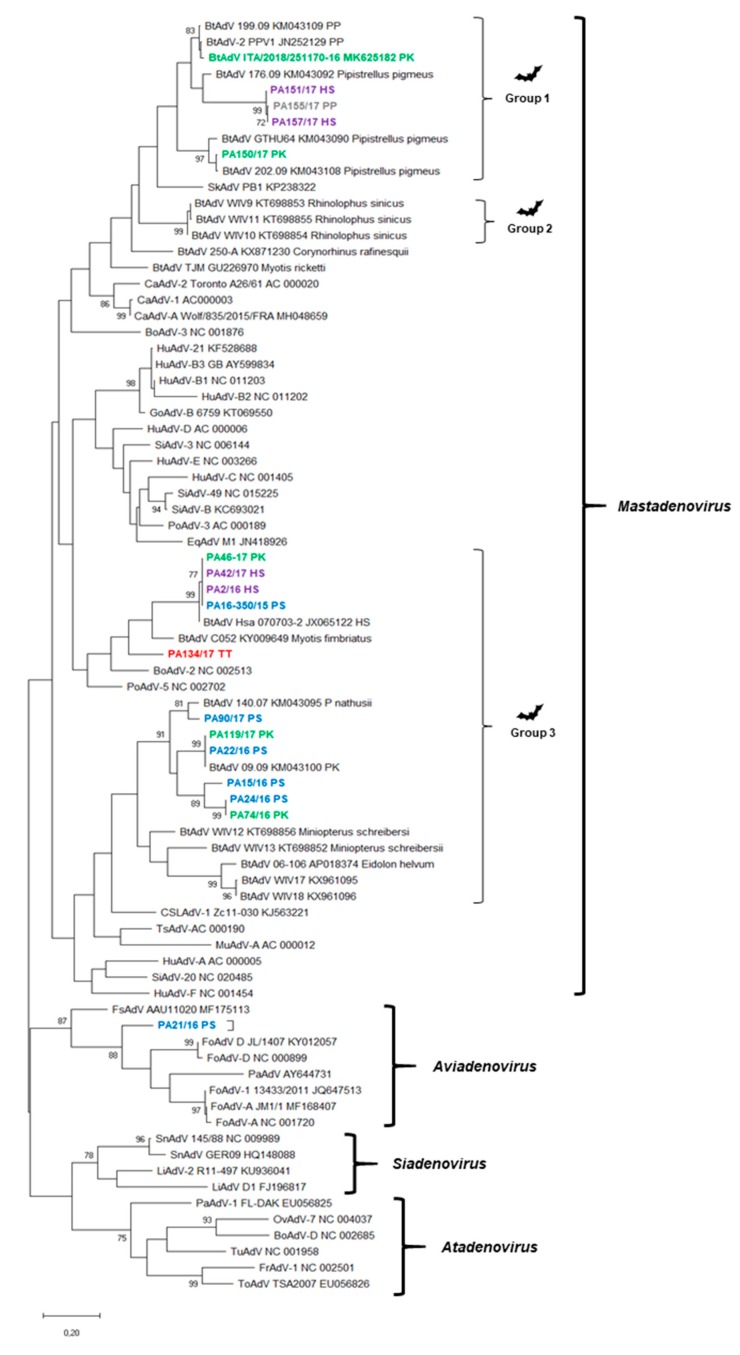
Adenoviruses are important pathogens of humans and animals. Bats have been recognized as potential reservoirs of novel viruses, with some viruses being regarded as a possible zoonotic threat to humans. In this study, we report the detection and analysis of adenoviruses from different bat species in northern Italy. Upon sequence and phylogenetic analysis, based on a short diagnostic fragment of the highly-conserved DNA polymerase gene, we identified potential novel candidate adenovirus species, including an avian-like adenovirus strain. An adenovirus isolate was obtained in simian cell lines from the carcass of a Pipistrellus kuhlii, and the complete genome sequence was reconstructed using deep sequencing technologies. The virus displayed high nucleotide identity and virtually the same genome organization as the Pipistrellus pipistrellus strain PPV1, isolated in Germany in 2007. Gathering data on epidemiology and the genetic diversity of bat adenoviruses may be helpful to better understand their evolution in the mammalian and avian hosts.

Keywords: Italy; NGS; adenovirus; aviadenovirus; bat; mastadenovirus; phylogenetic analysis; sequence.


Conflict of interest statement

The authors declare no conflict of interest.

Figures

Figure 1
Figure 1
Detection of adenovirus isolate ITA/2018/251170-16 with MARC-145 cells. (a) Control cells, 10x magnification; (b) Infected cells showing CPE, 10× magnification; (c) negative staining (NaPT 2%) electron microscopy of ITA/2018/251170-16 virion. Bar represents 200 nm.
Figure 2
Figure 2
Genomic organization of strain ITA/2018/251170-16. The genome is represented by a black horizontal line marked at 1000-bp intervals. The predicted ORFs are shown as arrows. Dashed lines represent potential splicing mechanisms.
Figure 3
Figure 3
Phylogenetic tree based on the full-length genome of representative members of Mastadenovirus, Aviadenovirus, Siadenovirus, and Atadenovirus genera. GenBank accession numbers are provided for reference strains. The tree was generated using the maximum-likelihood method with the Jukes–Cantor algorithm of distance correction, with bootstrapping up to 1000 replicates. Bootstrap values >70% are shown. Scale bar indicates nt substitutions per site. Black arrow indicates the strain retrieved in this study. CaAdV, canine adenovirus, BtAdV, bat adenovirus, SkAdV, skunk adenovirus, EqAdV, equine adenovirus, PoAdV, porcine adenovirus, BoAdV, bovine adenovirus, SiAdV, simian adenovirus, HuAdV, human adenovirus, TsAdV, tree shrew adenovirus, OvAdV, ovine adenovirus, MuAdV, murine adenovirus, CSLAdV, California sea lion adenovirus, FoAdV, fowl adenovirus, FrAdV, frog adenovirus, TuAdV, turkey adenovirus, SnAdV, snake adenovirus.
Figure 4
Figure 4
Phylogenetic tree based on the partial polymerase-coding region (233 nt positions) of adenoviruses. GenBank accession numbers are provided for reference strains. The tree was generated using the maximum-likelihood method with the Jukes–Cantor method by bootstrapping over 1000 replicates. Bootstrap values >70% are shown. Scale bar indicates nt substitutions per site. Blackarrows indicates the strains sequenced in this study. PS, Pipistrellus spp. (blue color); PP, Pipistrellus pipistrellus (grey color); PK, Pipistrellus kuhlii (green color); HS, Hypsugo savii (purple color); TT, Tadarida teniotis (red color). BtAdV, bat adenovirus, SkAdV, skunk adenovirus, EqAdV, equine adenovirus, CaAdV, canine adenovirus, HuAdV, human adenovirus, GoAdV, gorilla adenovirus, SiAdV, simian adenovirus, PoAdV, porcine adenovirus, MuAdV, murine adenovirus, TsAdV, tree shrew adenovirus, CSLAdV, California sea lion adenovirus, BoAdV, bovine adenovirus, FsAdV, fur seal adenovirus, PaAdV, parrot adenovirus, FoAdV, fowl adenovirus, SnAdV, snake adenovirus, LiAdV, lizard adenovirus, OvAdV, ovine adenovirus, TuAdV, turkey adenovirus, ToAdV, tortoise adenovirus, FrAdV, frog adenovirus.

References

    1. Harrach B., Benkö M., Both G., Brown M., Davison A., Echavarría M., Hess M., Jones M., Kajon A., Lehmkuhl H., et al. Family Adenoviridae. In: King A., Adams M., Carstens E., Lefkowitz E., editors. Virus Taxonomy: Classification and Nomenclature of Viruses: Ninth Report of the International Committee on Taxonomy of Viruses. Elsevier; San Diego, CA, USA: 2011. pp. 125–141.
    1. Tan B., Yang X.L., Ge X.Y., Peng C., Liu H.Z., Zhang Y.Z., Zhang L.B., Shi Z.L. Novel bat adenoviruses with low G+C content shed new light on the evolution of adenoviruses. J. Gen. Virol. 2017;98:739–748. doi: 10.1099/jgv.0.000739. - DOI - PubMed
    1. Davison A.J., Benko M., Harrach B. Genetic content and evolution of adenoviruses. J. Gen. Virol. 2003;84:2895–2908. doi: 10.1099/vir.0.19497-0. - DOI - PubMed
    1. International Committee on Taxonomy of Viruses (ICTV) Virus Taxonomy: 2018b Release. [(accessed on 2 April 2019)]; Available online: http://talk.ictvonline.org/taxonomy/
    1. Pauly M., Hoppe E., Mugisha L., Petrzelkova K., Akoua-Koffi C., Couacy-Hymann E., Anoh A.E., Mossoun A., Schubert G., Wiersma L., et al. High prevalence and diversity of species D adenoviruses (HAdV-D) in human populations of four Sub-Saharan countries. Virol. J. 2014;11:25. doi: 10.1186/1743-422X-11-25. - DOI - PMC - PubMed

Publication types

LinkOut - more resources