. 2019 Jun 7;18(1):72.
doi: 10.1186/s12938-019-0690-x.

Prediction of the mechanical response of cardiac alternans by using an electromechanical model of human ventricular myocytes

Affiliations

Prediction of the mechanical response of cardiac alternans by using an electromechanical model of human ventricular myocytes

Jun Ik Park et al. Biomed Eng Online. .

Abstract

Purpose: Although the quantitative analysis of electromechanical alternans is important, previous studies have focused on electrical alternans, and there is a lack quantitative analysis of mechanical alternans at the subcellular level according to various basic cycle lengths (BCLs). Therefore, we used the excitation-contraction (E-C) coupling model of human ventricular cells to quantitatively analyze the mechanical alternans of ventricular cells according to various BCLs.

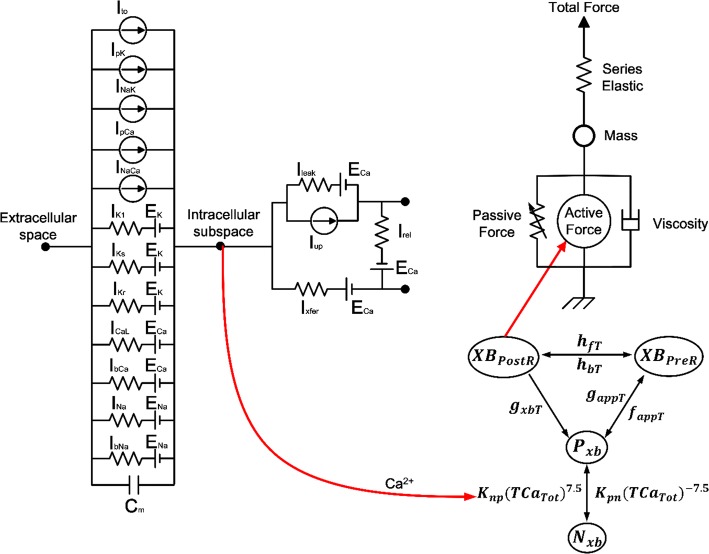
Methods: To implement E-C coupling, we used calcium transient data, which is the output data of electrical simulation using the electrophysiological model of human ventricular myocytes, as the input data of mechanical simulation using the contractile myofilament dynamics model. Moreover, we applied various loads on ventricular cells for implementation of isotonic and isometric contraction.

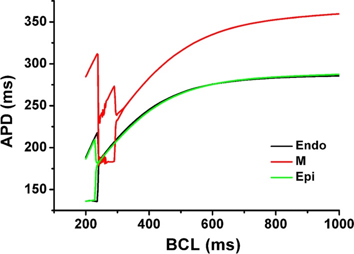
Results: As the BCL was reduced from 1000 to 200 ms at 30 ms increments, mechanical alternans, as well as electrical alternans, were observed. At this time, the myocardial diastolic tension increased, and the contractile ATP consumption rate remained greater than zero even in the resting state. Furthermore, the time of peak tension, equivalent cell length, and contractile ATP consumption rate were all reduced. There are two tendencies that endocardial, mid-myocardial, and epicardial cells have the maximum amplitude of tension and the peak systolic tension begins to appear at a high rate under the isometric condition at a particular BCL.

Conclusions: We observed mechanical alternans of ventricular myocytes as well as electrical alternans, and identified unstable conditions associated with mechanical alternans. We also determined the amount of BCL given to each ventricular cell to generate stable and high tension state in the case of isometric contraction.

Keywords: Alternans; Basic cycle length; Excitation–contraction coupling model; Human ventricular myocyte; Simulation study.


Conflict of interest statement

The authors declare that they have no competing interests.

Figures

Fig. 1
Fig. 1
Schematic diagram of excitation–contraction coupling model of ventricular cell. The left diagram represents a human ventricular cell model with electrophysiological characteristics that mimic the ion exchange phenomenon through the cell membrane of myocytes. Ito is the transient outward K+ current, IpK is the plateau K+ pump current, INaK is the Na+–K+ ion exchanger current, IpCa is plateau Ca2+ pump current, and INaCa is the Na+–Ca2+ ion exchanger current. Ek, ECa, and ENa are the equilibrium potentials of K+, Ca2+, and Na+ ions, respectively. Cm is the ventricular cell membrane capacitance in the unit surface area. IK1 is the inward rectifier K1 current, IKs is the slow delayed rectifier K+ current, IK1 is the rapid delayed rectifier K+ current, ICaL is the L-type inward Ca2+ current, IbCa denotes the background Ca2+ current, IbNa is the background Na+ current, and INa is the fast inward Na+ current. Irel is the release Ca2+ current from the sarcoplasmic reticulum (SR), Ileak is the leakage Ca2+ current from the SR, and Iup is the Ca2+ uptake current in the SR. The right diagram represents the cardiac myofilament model to simulate mechanical responses of myocytes. Nxb and Pxb are non-permissive and permissive confirmations of regulatory proteins, respectively, and XBPreR and XBPostR represent the probability that the cross-bridge is in the pre/post-rotated force-generating state. gxbT is the detachment transition rate with consuming ATP, hfT and hbT are the forward and backward transition rates, fappT and gaapT are the cross-bridge attachment rate of transition and reverse rate. Knp and Kpn are transition rates for the fraction of permissive, Knp(TCaTot)7.5 is the forward rate of the non-permissive to permissive transition in the opposite direction, and Kpn(TCaTot)− 7.5 is the backward rate of the permissive to non-permissive transition. There are two types forces: active force and passive force. The active force created by contraction of the cross-bridge, and the passive force improves the complete muscle response with titin and other cytoskeletal elements. Mass prevents prompt changes in muscle-shortening velocity for quick-release protocols. Series elastic element represents effects of compliant end connections on real muscle preparations
Fig. 2
Fig. 2
Changes in action potential duration (APD) according to basic cycle length (BCL) for each ventricular cell. Endo is the endocardial cell, M is the mid-myocardial cell, and Epi is the epicardial cell
Fig. 3
Fig. 3
Electrical simulations of membrane potential and intracellular calcium concentration. ac Cases of Endo, M, and Epi, respectively for 1 s. Each figure is divided into three cases according to different BCLs (1000 ms, 280 ms when electrical alternans occur under Endo and Epi conditions; and 200 ms when electrical alternans appear under M condition)
Fig. 4
Fig. 4
Mechanical simulations of myocardial tension and contractile ATP consumption rate. The left side represents the results of isometric contraction when the load is 1000 kPa (mN/mm2) and the right side shows isotonic contraction when the load is 10 kPa
Fig. 5
Fig. 5
Mechanical simulations of the change in equivalent cell length in the case of three different BCLs (left) and total time from BCL of 1000 ms to 200 ms (right) when the load applied to each ventricular cell is 10 kPa and 0.6 kPa. On the right side, arrow indicates the specific BCL which takes place at the point in which the diastolic equivalent cell length is not relaxed to 1.0 (100%)
Fig. 6
Fig. 6
Mechanical simulations of amplitude and systolic peak of myocardial tension according to decreasing of BCL under isometric condition

References

    1. Banville I, Gray RA. Effect of APD and CV restitution and their spatial dispersion on alternans and the stability of arrhythmias. J Cardiovasc Electrophysiol. 2002;13:1141–1149. doi: 10.1046/j.1540-8167.2002.01141.x. - DOI - PubMed
    1. Cherry EM, Fenton FH. Suppression of alternans and conduction blocks despite steep APD restitution: electrotonic, memory, and conduction velocity restitution effects. Am J Physiol Circ Physiol. 2004;286:H2332–H2341. doi: 10.1152/ajpheart.00747.2003. - DOI - PubMed
    1. Watanabe MA, Fenton FH, Evans SJ, Hastings HM, Karma A. Mechanisms for discordant alternans. J Cardiovasc Electrophysiol. 2001;12:196–206. doi: 10.1046/j.1540-8167.2001.00196.x. - DOI - PubMed
    1. Pastore JM, Girouard SD, Laurita KR, Akar FG, Rosenbaum DS. Mechanism linking T-wave alternans to the genesis of cardiac fibrillation. Circulation. 1999;99:1385–1394. doi: 10.1161/01.CIR.99.10.1385. - DOI - PubMed
    1. Shiferaw Y, Karma A. Turing instability mediated by voltage and calcium diffusion in paced cardiac cells. Proc Natl Acad Sci. 2006;103:5670–5675. doi: 10.1073/pnas.0511061103. - DOI - PMC - PubMed