Review
. 2019 Jun 7;23(1):209.
doi: 10.1186/s13054-019-2490-x.

Decompressive craniectomy for acute ischemic stroke

Affiliations
Review

Decompressive craniectomy for acute ischemic stroke

Thomas Beez et al. Crit Care. .

Abstract

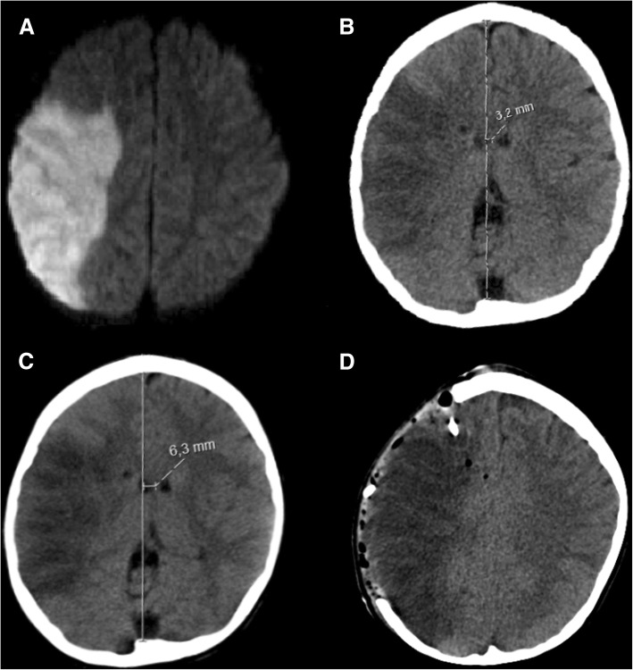
Malignant stroke occurs in a subgroup of patients suffering from ischemic cerebral infarction and is characterized by neurological deterioration due to progressive edema, raised intracranial pressure, and cerebral herniation. Decompressive craniectomy (DC) is a surgical technique aiming to open the "closed box" represented by the non-expandable skull in cases of refractory intracranial hypertension. It is a valuable modality in the armamentarium to treat patients with malignant stroke: the life-saving effect has been proven for both supratentorial and infratentorial DC in virtually all age groups. This leaves physicians with the difficult task to decide who will require early or preemptive surgery and who might benefit from postponing surgery until clear evidence of deterioration evolves. Together with the patient's relatives, physicians also have to ascertain whether the patient will have acceptable disability and quality of life in his or her presumed perception, based on preoperative predictions. This complex decision-making process can only be managed with interdisciplinary efforts and should be supported by continued research in the age of personalized medicine.

Keywords: Cranioplasty; Decompressive craniectomy; Hemicraniectomy; Malignant ischemic infarction; Pediatric stroke; Suboccipital craniectomy.


Conflict of interest statement

The authors declare that they have no competing interests.

Figures

Fig. 1
Fig. 1
Stepwise reduction of ICP during DC. Representative ICP measurements obtained during DC performed on an 11-year-old boy suffering from refractory intracranial hypertension. Removal of the bone flap reduces ICP by 66% from 30 to 10 mmHg, followed by a further 50% reduction to 5 mmHg after dural opening
Fig. 2
Fig. 2
Decompressive hemicraniectomy for malignant ischemic stroke. Axial CT scan before surgery (a), demonstrating a demarcated right-sided MCA infarct (highlighted in red) with hemorrhagic transformation (black arrow) and midline shift to the left side (red line). Axial CT scan after surgery (b), showing the craniectomy defect (highlighted in green) with decompressed lateral ventricle (highlighted in blue) and reversal of midline shift (green line)
Fig. 3
Fig. 3
Operative technique of supratentorial DC. Artist’s rendition of a human head (a) with a typical incision line for DC (gray line). 3D reconstruction of a human skull (b) demonstrating burr holes (gray circles), craniectomy (gray area), and additional osteoclastic decompression of the middle cranial fossa floor (hatched area) as well as typical dural incision (red lines). 3D reconstruction of a human skull (c) with a typical hemicraniectomy skull defect. Intraoperative photography of a human brain after DC (d)
Fig. 4
Fig. 4
Suboccipital decompressive craniectomy for malignant cerebellar stroke. Axial CT scan before surgery (a), showing a large demarcated cerebellar infarct (highlighted in red). Axial CT scan after surgery (b), demonstrating the craniectomy defect (highlighted in green) and decompressed fourth ventricle (highlighted in blue)
Fig. 5
Fig. 5
Operative technique of infratentorial DC. Artist’s rendition of a human head (a) with a typical incision line for suboccipital DC (gray line). 3D reconstruction of a human skull (b) demonstrating burr holes (gray circles), craniectomy, and removal of the posterior arch of the atlas (gray areas) as well as typical dural incision (red lines). 3D reconstruction of a human skull (c) with a typical suboccipital DC skull defect
Fig. 6
Fig. 6
Cranioplasty and autologous bone flap resorption. 3D reconstruction of a 57-year-old male patient’s skull after autologous cranioplasty following DC for left-sided malignant MCA infarction (a). One year later (b), significant areas of the bone flap resorption occurred (highlighted in red). The explanted autologous bone flap (c) shows the overall thinning and obvious holes due to resorption. A typical example of an alloplastic implant (d) after right-sided DC in another patient (11-year-old boy after TBI)
Fig. 7
Fig. 7
Mortality at 12 months after malignant MCA infarction. Forest plot presenting risk difference and 95% confidence interval (CI) for a pooled analysis of mortality at 12 months from RCTs comparing DC and best medical care
Fig. 8
Fig. 8
Hygroma occurring after DC. Axial CT scan showing ipsilateral and contralateral subdural hygroma (black arrows), which occurred after left-sided hemicraniectomy. Additionally, a large epidural fluid collection (white arrow) is observed
Fig. 9
Fig. 9
Sunken skin flap after DC. Axial CT scan showing a sunken skin flap (white arrow) after left-sided hemicraniectomy
Fig. 10
Fig. 10
Pediatric ischemic stroke. A representative example of pediatric ischemic stroke in a 6-year-old boy with sickle cell anemia: axial diffusion-weighted MRI sequence (a) with increased signal in the right MCA territory, indicating ischemic infarction. Axial CT scan obtained on day 1 after stroke onset (b), showing a demarcated infarct with 3.2 mm midline shift. Axial CT scan obtained on day 2 after stroke onset (c), revealing a progressive midline shift of 6.3 mm, correlating with neurological deterioration. Axial CT scan after hemicraniectomy and implantation of a right frontal intraparenchymal ICP probe (d), with reversal of midline shift

References

    1. Goodrich JT. How to get in and out of the skull: from tumi to “hammer and chisel” to the Gigli saw and the osteoplastic flap. Neurosurg Focus. 2014;36:E6. doi: 10.3171/2014.2.FOCUS13543. - DOI - PubMed
    1. Kocher T. Hirnerschütterung, Hirndruck und chirurgische Eingriffe bei Hirnkrankheiten. Wien: Alfred Hölder; 1901.
    1. Cushing HI. Subtemporal decompressive operations for the intracranial complications associated with bursting fractures of the skull. Ann Surg. 1908;47:641–644.1. doi: 10.1097/00000658-190805000-00001. - DOI - PMC - PubMed
    1. Scarcella G. Encephalomalacia simulating the clinical and radiological aspects of brain tumor; a report of 6 cases. J. Neurosurg. 1956;13:278–292. doi: 10.3171/jns.1956.13.4.0278. - DOI - PubMed
    1. King AB. Massive cerebral infarction producing ventriculographic changes suggesting a brain tumor. J Neurosurg. 1951;8:536–539. doi: 10.3171/jns.1951.8.5.0536. - DOI - PubMed