. 2019 Jun 7;7(1):91.
doi: 10.1186/s40478-019-0743-1.

Resistance and resilience to Alzheimer's disease pathology are associated with reduced cortical pTau and absence of limbic-predominant age-related TDP-43 encephalopathy in a community-based cohort

Affiliations

Resistance and resilience to Alzheimer's disease pathology are associated with reduced cortical pTau and absence of limbic-predominant age-related TDP-43 encephalopathy in a community-based cohort

Caitlin S Latimer et al. Acta Neuropathol Commun. .

Erratum in

Abstract

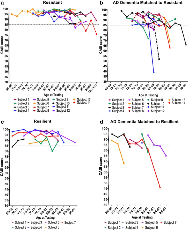
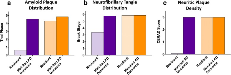
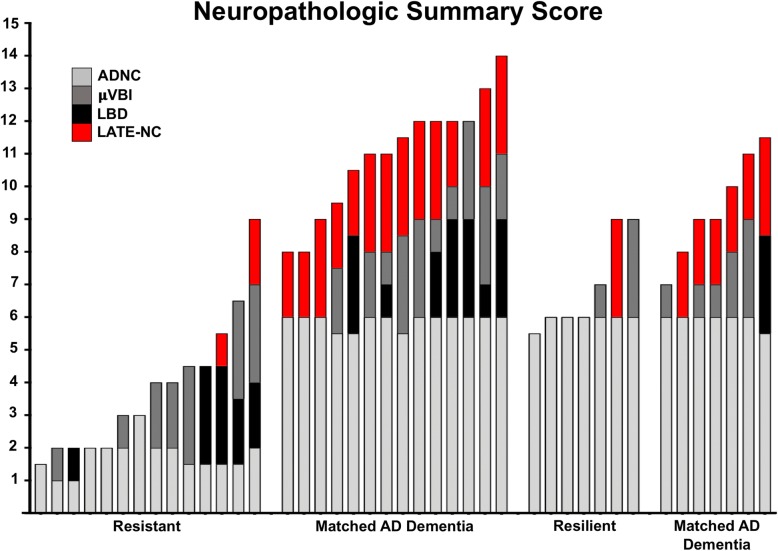
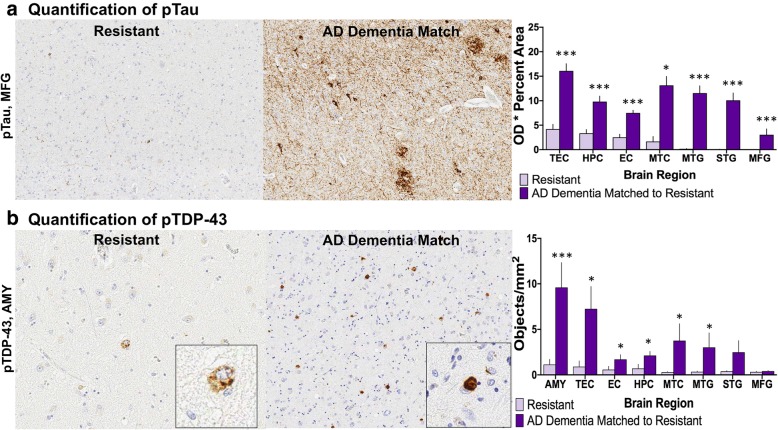
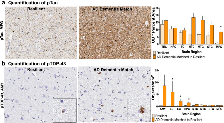
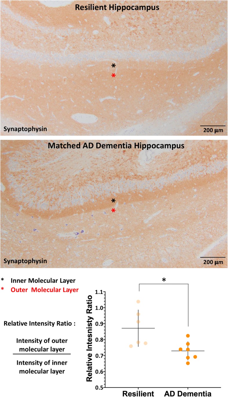
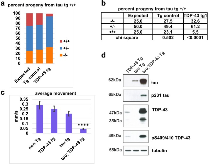
Alzheimer's disease neuropathologic change (ADNC) is defined by progressive accumulation of β-amyloid plaques and hyperphosphorylated tau (pTau) neurofibrillary tangles across diverse regions of brain. Non-demented individuals who reach advanced age without significant ADNC are considered to be resistant to AD, while those burdened with ADNC are considered to be resilient. Understanding mechanisms underlying ADNC resistance and resilience may provide important clues to treating and/or preventing AD associated dementia. ADNC criteria for resistance and resilience are not well-defined, so we developed stringent pathologic cutoffs for non-demented subjects to eliminate cases of borderline pathology. We identified 14 resistant (85+ years old, non-demented, Braak stage ≤ III, CERAD absent) and 7 resilient (non-demented, Braak stage VI, CERAD frequent) individuals out of 684 autopsies from the Adult Changes in Thought study, a long-standing community-based cohort. We matched each resistant or resilient subject to a subject with dementia and severe ADNC (Braak stage VI, CERAD frequent) by age, sex, year of death, and post-mortem interval. We expanded the neuropathologic evaluation to include quantitative approaches to assess neuropathology and found that resilient participants had lower neocortical pTau burden despite fulfilling criteria for Braak stage VI. Moreover, limbic-predominant age-related TDP-43 encephalopathy neuropathologic change (LATE-NC) was robustly associated with clinical dementia and was more prevalent in cases with high pTau burden, supporting the notion that resilience to ADNC may depend, in part, on resistance to pTDP-43 pathology. To probe for interactions between tau and TDP-43, we developed a C. elegans model of combined human (h) Tau and TDP-43 proteotoxicity, which exhibited a severe degenerative phenotype most compatible with a synergistic, rather than simply additive, interaction between hTau and hTDP-43 neurodegeneration. Pathways that underlie this synergy may present novel therapeutic targets for the prevention and treatment of AD.

Keywords: Alzheimer’s disease neuropathologic change; C. elegans; Dementia; Hyperphosphorylated tau; Resilience; Resistance; TDP-43.


Conflict of interest statement

The authors declare that they have no competing interests.

Figures

Fig. 1
Fig. 1
Longitudinal Cognitive Scores. Resistant (a) and resilient (c) subjects show relatively stable cognitive performance over time. The AD dementia matched subjects (b and d) were followed until the subjects came to consensus conference (when CASI ≤ 85) and a diagnosis of dementia was made, after which most subjects are no longer administered cognitive testing. Matched pairs are denoted by color and line style
Fig. 2
Fig. 2
AD Neuropathologic change. Cases were selected based on the degree of AD neuropathologic change. The resistant cases were selected based on a Braak score of III or less and absent neuritic plaques by CERAD criteria. Their AD dementia matches, the resilient cases, and their respective AD dementia matches were selected based on a Braak stage VI and frequent neuritic plaques. Re-evaluation demonstrated the expected distribution of amyloid plaques (a), neurofibrillary tangles (b), and density of neuritic plaques (c), based on these selection criteria
Fig. 3
Fig. 3
Neuropathology Summary Score. Every subject is depicted by a bar, which is subdivided by colors to represent the different types of neuropathology present for each subject. The grey bars represent AD neuropathologic change (ADNC) and are calculated as (Braak stage/2) + CERAD score, such that the maximum score for ADNC is 6. The slate bars represent microvascular brain injury (μVBI), specifically the microinfarct burden, such that 1 microinfarct = 1, 2 microinfarcts = 2, and 3 or more microinfarcts = 3. The black bars represent extent of Lewy body disease (LBD), such that 1 = brainstem only, 2 = limbic/amygdala predominant, 3 = neocortical. The red bars represent limbic-predominant age-related TDP-43 neuropathologic change (LATE-NC), such that 1 = amygdala only, 2 = hippocampal, and 3 = neocortical (beyond medial temporal). Overall, the resistant and resilient groups conspicuously lack LATE-NC, while pTDP-43 pathology is nearly ubiquitously present in subjects with dementia
Fig. 4
Fig. 4
Semi-quantification of pathologic proteins. The burden of each pathologic protein (Aβ, pTau, and pTDP-43) was assessed in each subject by assigning a score of 0–3 for Aβ, 0–3 for pTau and 0–6 for pTDP-43. a As expected (based on selection criteria), the resistant group had very little pathology resulting in significant differences for each pathologic protein in nearly every brain region assessed compared to the AD dementia matches. b The resilient group showed no significant differences in burden of Aβ pathology throughout the cortex, but did show significantly lower Aβ in midbrain and cerebellum. pTau pathology was significantly less only in the MFG in resilient cases. pTDP-43 pathology showed significantly less pathologic burden in the AMY and HPC. (MFG, middle frontal gyrus, IPL, inferior parietal lobule; MTG, middle temporal gyrus; STG, superior temporal gyrus; OC, occipital cortex; STR, striatum; MB, midbrain; CBL, cerebellum; HPC, hippocampus; AMY, amygdala) ***p < 0.005, **p < 0.01, *p < 0.05; Wilcoxon matched-pairs signed-ranks test
Fig. 5
Fig. 5
Cortical microvacuolar change. The degree of neurodegenerative tissue damage was assessed in each case on H&E/LFB-stained slides. The microvacuolar changes associated with parenchymal loss and reactive gliosis were scored on a 3-point scale in each neocortical region such that 1 = limited to superficial layers (1–2), 2 = extends to deeper layers (3–4), and 3 = translaminar involvement (5–6). In all AD dementia subjects, the MTG was the most severely affected. a Overall there was significantly less parenchymal damage in the resistant group in the IPL, MTG, and OC. There was a trend for less parenchymal damage in the MTG (p = 0.0588) and the STG (p = 0.0592). b Resilient subjects compared to AD dementia matches showed less parenchymal damage in all cortical regions assessed. (MFG, middle frontal gyrus; IPL, inferior parietal lobule; MTG, middle temporal gyrus; STG, superior temporal gyrus; and OCX, occipital cortex). *p < 0.05; Wilcoxon matched-pairs signed-ranks test
Fig. 6
Fig. 6
Quantitative pathology for pTau and pTDP-43 in the resistant group. The resistant group had less quantitative pTau in every brain region assessed (a) and less quantitative pTDP-43 burden in the majority of brain regions assessed (b) compared to the AD dementia matches. (MFG, middle frontal gyrus; MTG, middle temporal gyrus; STG, superior temporal gyrus; MTC, mesial temporal cortex; EC, entorhinal cortex; HPC, hippocampus; TEC, transentorhinal cortex; AMY, amygdala) ***p < 0.005, *p < 0.05; Wilcoxon matched-pairs signed-ranks test
Fig. 7
Fig. 7
Quantitative pathology for pTau and pTDP-43 in the resilient group: a The resilient group had less quantitative pTau burden in the MFG. b Quantitative assessments of pTDP-43 also revealed less pathologic burden in TEC, EC, and HPC in the resilient group compared to AD dementia matches. Amygdala showed a trend for reduced TDP-43 immunoreactivity (p = 0.0630). (MFG, middle frontal gyrus; MTG, middle temporal gyrus; STG, superior temporal gyrus; MTC, mesial temporal cortex; EC, entorhinal cortex; HPC, hippocampus; TEC, transentorhinal cortex; AMY, amygdala) *p < 0.05; Wilcoxon matched-pairs signed-ranks test
Fig. 8
Fig. 8
Synaptic integrity of the perforant pathway in the resilient group. The ratio of synaptophysin staining between the outer molecular layer of the dentate gyrus of the hippocampus (red asterisk) and the inner molecular layer (black asterisk) provides a measure of synaptic integrity of the perforant pathway. The AD dementia subjects had a reduced ratio due to a preferential reduction in staining of the outer molecular layer compared to the inner layer while the resilient group maintained a ratio closer to one, reflecting maintained synaptic health. *p < 0.05, Wilcoxon matched-pairs signed-ranks test
Fig. 9
Fig. 9
hTau and hTDP-43 synergize in vivo to drive neurotoxicity and protein accumulation. a, b Pan-neuronal expression of human TDP-43 (hTDP-43) is synthetic lethal with human Tau (hTau) in C. elegans transgenic models. Progeny from animals homozygous for hTau tg (+/+) but heterozygous for either GFP Tg or hTDP-43 tg (+/−) were picked blind and then scored for GFP Tg or hTDP-43 tg genotype. Expected Mendelian ratios for assortment of a single genetic element are 25% (+/+), 50% (+/−), and 25% (−/−). Ratios of progeny from animals heterozygous for GFP Tg are not significantly different from Mendelian ratios (p = 0.502, Chi square analysis). Ratios of progeny from animals heterozygous for hTDP-43 tg are significantly different from Mendelian ratios (p < 0.0001). N = 563, GFP Tg. N = 561, hTDP-43 tg. c Developmentally synchronized L4 larvae of hTau tg (+/+); TDP-43 (+/+) double homozygotes move significantly less than hTau tg (+/+) or hTDP-43 tg (+/+) alone (****p < 0.0005). Statistical significance was determined using one-way ANOVA with Tukey’s multiple-comparison test. d Co-expression of hTau and hTDP-43 promotes accumulation and pathological phosphorylation of both proteins in vivo. Developmentally synchronized day 1 adult C. elegans were harvested and tested by immunoblot for total tau, phosphorylated tau (AT180), total TDP-43, phosphorylated TDP-43 (phospho-S409/410) and tubulin (load control). Immunoblot shown is representative of three independent replicate experiments

References

    1. Abner EL, Kryscio RJ, Schmitt FA, Santacruz KS, Jicha GA, Lin Y, Neltner JM, Smith CD, Van Eldik LJ, Nelson PT. “End-stage” neurofibrillary tangle pathology in preclinical Alzheimer’s disease: fact or fiction? J Alzheimers Dis. 2011;25:445–453. doi: 10.3233/JAD-2011-101980. - DOI - PMC - PubMed
    1. Amador-Ortiz C, Lin W-L, Ahmed Z, Personett D, Davies P, Duara R, Graff-Radford NR, Hutton ML, Dickson DW. TDP-43 immunoreactivity in hippocampal sclerosis and Alzheimer’s disease. Ann Neurol. 2007;61:435–445. doi: 10.1002/ana.21154. - DOI - PMC - PubMed
    1. American Psychiatric Association. Task force on DSM-IV. (2000) Diagnostic and statistical manual of mental disorders: DSM-II. Am Psychiatr Publ. doi: http://dx.doi.org.lrc1.usuhs.edu/10.1176/appi.books.9780890425596 - DOI
    1. Aoki Naoya, Murray Melissa E., Ogaki Kotaro, Fujioka Shinsuke, Rutherford Nicola J., Rademakers Rosa, Ross Owen A., Dickson Dennis W. Hippocampal sclerosis in Lewy body disease is a TDP-43 proteinopathy similar to FTLD-TDP Type A. Acta Neuropathologica. 2014;129(1):53–64. doi: 10.1007/s00401-014-1358-z. - DOI - PMC - PubMed
    1. Arenaza-Urquijo EM, Vemuri P. Resistance vs resilience to Alzheimer disease: clarifying terminology for preclinical studies. Neurology. 2018;90:695–703. doi: 10.1212/WNL.0000000000005303. - DOI - PMC - PubMed

Publication types

MeSH terms