

Retinal Microvascular and Neurodegenerative Changes in Alzheimer's Disease and Mild Cognitive Impairment Compared with Control Participants
- PMID: 31174670
- PMCID: PMC6586560
- DOI: 10.1016/j.oret.2019.02.002
Retinal Microvascular and Neurodegenerative Changes in Alzheimer's Disease and Mild Cognitive Impairment Compared with Control Participants
Abstract
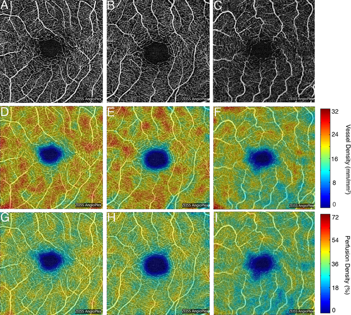
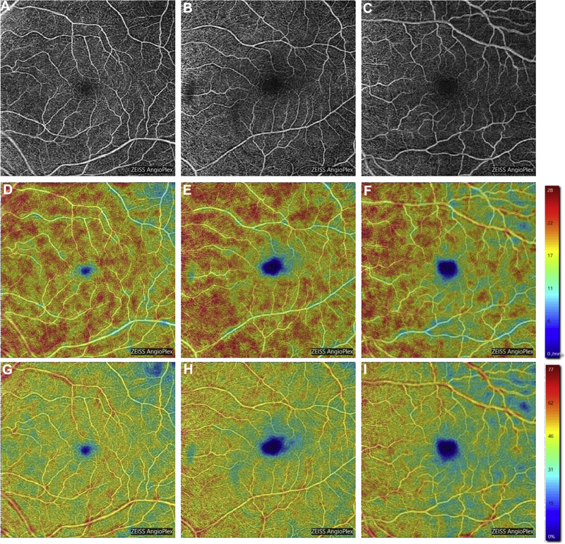
Purpose: Evaluate and compare the retinal microvasculature in the superficial capillary plexus (SCP) in Alzheimer's disease (AD), mild cognitive impairment (MCI), and cognitively intact controls using OCT angiography. OCT parameters were also compared.
Design: Cross-sectional study.
Participants: Seventy eyes from 39 AD participants, 72 eyes from 37 MCI participants, and 254 eyes from 133 control participants were enrolled.
Methods: Participants were imaged using Zeiss Cirrus HD-5000 with AngioPlex (Carl Zeiss Meditec, Dublin, CA) and underwent cognitive evaluation with Mini-Mental State Examination.
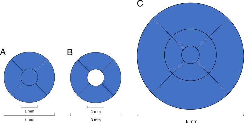
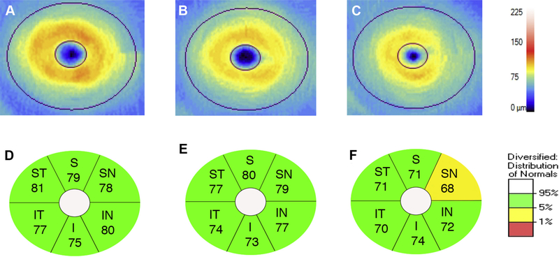
Main outcome measures: Vessel density (VD) and perfusion density (PD) in the SCP within the Early Treatment Diabetic Retinopathy Study 6-mm circle, 3-mm circle, and 3-mm ring were compared between groups. Foveal avascular zone (FAZ) area, central subfield thickness (CST), macular ganglion cell-inner plexiform layer (GC-IPL) thickness, and peripapillary retinal nerve fiber layer (RNFL) thickness were also compared.
Results: Alzheimer's participants showed significantly decreased SCP VD and PD in the 3-mm ring (P = 0.001 and P = 0.002, respectively) and 3-mm circle (P = 0.003 and P = 0.004, respectively) and decreased SCP VD in the 6-mm circle (P = 0.047) compared with MCI and significantly decreased SCP VD and PD in the 3-mm ring (P = 0.008 and P = 0.004, respectively) and 3-mm circle (P = 0.015 and P = 0.009, respectively) and SCP PD in the 6-mm circle (P = 0.033) when compared with cognitively intact controls. There was no difference in SCP VD or PD between MCI and controls (P > 0.05). FAZ area and CST did not differ significantly between groups (P > 0.05). Alzheimer's participants showed significantly decreased GC-IPL thickness over the inferior (P = 0.032) and inferonasal (P = 0.025) sectors compared with MCI and significantly decreased GC-IPL thickness over the entire (P = 0.012), superonasal (P = 0.041), inferior (P = 0.004), and inferonasal (P = 0.006) sectors compared to controls. MCI participants showed significantly decreased temporal RNFL thickness (P = 0.04) compared with controls.
Conclusions: Alzheimer's participants showed significantly reduced macular VD, PD, and GC-IPL thickness compared with MCI and controls. Changes in the retinal microvasculature may mirror small vessel cerebrovascular changes in AD.
Copyright © 2019 American Academy of Ophthalmology. Published by Elsevier Inc. All rights reserved.
Conflict of interest statement
Figures
References
-
- Galton CJ, Patterson K, Xuereb JH, Hodges JR. Atypical and typical presentations of Alzheimer’s disease: a clinical, neuropsychological, neuroimaging and pathological study of 13 cases. Brain 2000;123:484–498. - PubMed
-
- Gauthier S, Reisberg B, Zaudig M, et al. Mild cognitive impairment. Lancet 2006;367:1262–1270. - PubMed
Publication types
MeSH terms
Grants and funding
LinkOut - more resources
Full Text Sources
Medical
Miscellaneous
