. 2019 Jul 17;103(2):203-216.e8.
doi: 10.1016/j.neuron.2019.05.013. Epub 2019 Jun 4.

Regional Heterogeneity in Gene Expression, Regulation, and Coherence in the Frontal Cortex and Hippocampus across Development and Schizophrenia

Affiliations

Regional Heterogeneity in Gene Expression, Regulation, and Coherence in the Frontal Cortex and Hippocampus across Development and Schizophrenia

Leonardo Collado-Torres et al. Neuron. .

Abstract

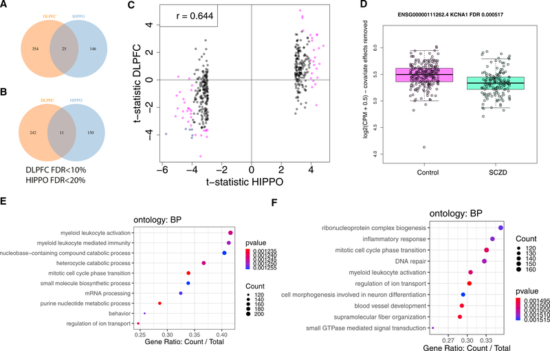
The hippocampus formation, although prominently implicated in schizophrenia pathogenesis, has been overlooked in large-scale genomics efforts in the schizophrenic brain. We performed RNA-seq in hippocampi and dorsolateral prefrontal cortices (DLPFCs) from 551 individuals (286 with schizophrenia). We identified substantial regional differences in gene expression and found widespread developmental differences that were independent of cellular composition. We identified 48 and 245 differentially expressed genes (DEGs) associated with schizophrenia within the hippocampus and DLPFC, with little overlap between the brain regions. 124 of 163 (76.6%) of schizophrenia GWAS risk loci contained eQTLs in any region. Transcriptome-wide association studies in each region identified many novel schizophrenia risk features that were brain region-specific. Last, we identified potential molecular correlates of in vivo evidence of altered prefrontal-hippocampal functional coherence in schizophrenia. These results underscore the complexity and regional heterogeneity of the transcriptional correlates of schizophrenia and offer new insights into potentially causative biology.

Keywords: RNA-seq; TWAS; brain; development; eQTL; human; regional coherence; schizophrenia.


Conflict of interest statement

DECLARATION OF INTERESTS

The following BrainSeq Consortium members have competing interests. M.M., T.S., K.T., and D.J.H. are employees of Astellas Pharma. N.J.B. and A.J.C. are employees of AstraZeneca. D.A.C., J.N.C., C.L.A.R., B.J.E., P.J.E., D.C.A., Y. Li, Y. Liu, K.M., B.B.M., J.E.S., and H.W. are employees of Eli Lilly and Company. M.F., D.H., and H.K. are employees of Janssen Research & Development LLC and Johnson and Johnson. M.D. and L.F. are employees of H. Lundbeck A/S. T.K.-T. and D.M. are employees of F. Hoffmann-La Roche. P.O., S.X., and J.Q. are former employees of Pfizer.

Figures

Figure 1.
Figure 1.. Adult and Prenatal DLPFC and HIPPO Expression Differences
(A) Replication rate with consistent fold change for the adult and prenatal differences between brain regions using the BrainSpan dataset (n = 16 for adult, n = 36 for prenatal) across Bonferroni p value thresholds for the exon, gene, and exon-exon junction features. The labels for each point denote the number of features considered at the given Bonferroni p value threshold. (B) Volcano plots of the differential expression signal between brain regions for each feature type and age group stratified by whether the differential expression replicated in BrainSpan (p < 0.05 and consistent fold change). (C) Differentially expressed features grouped by gene ID for the adult and prenatal age groups. See also Figures S5–S10 and Tables S11 and S14.
Figure 2.
Figure 2.. Differences across Development between the DLPFC and HIPPO
(A) Estimated cell type RNA fractions for fetal quiescent neurons (FQNs), neurons, and oligodendrocytes. See Figure S11A for the other five cell type RNA fractions estimated with RNA deconvolution. (B) Differentially expressed features between brain regions across development grouped by gene ID. (C) GABRD is differentially expressed between the DLPFC and HIPPO across development (Bonferroni p value < 1 × 10⁻²²). The y axis shows the residualized expression after removing modeled covariates. PCW, post-conception week. See also Figures S11–S19 and Tables S1, S11, and S14.
Figure 3.
Figure 3.. SCZD Differential Expression in the DLPFC and HIPPO
(A) Venn diagram of differentially expressed genes with higher expression in neurotypical controls than SCZD-affected individuals by brain region (DLPFC, FDR < 10%; HIPPO, FDR < 20%). (B) Similar to (A) but for genes with higher expression in SCZD than in neurotypical controls. (C) t-statistics for the top 400 differentially expressed genes in the HIPPO compared with their t-statistics in the DLPFC. Pink points, FDR < 5% in the DLPFC or HIPPO; blue points, FDR < 5% in both brain regions. (D) KCNA1 is differentially expressed in the DLPFC (FDR = 0.000517). (E and F) Gene set enrichment analysis (GSEA) on the DLPFC (E) with biological process ontology terms and HIPPO (F). Count, number of enriched genes for the given ontology term; gene ratio, ratio of genes enriched for the ontology term among the number of genes that are contained in the given ontology term. Terms are colored by their p value of enrichment. See also Figures S24 and S26–S35 and Tables S2, S4, S5, S11, and S14.
Figure 4.
Figure 4.. DLPFC and HIPPO eQTLs and Schizophrenia Risk
(A) HIPPO eQTLs grouped by gene ID. (B) Region-dependent eQTLs grouped by SNP ID. (C) Region-dependent eQTLs group by gene ID. (D) Unique risk schizophrenia GWAS index SNPs from PCG2 (Pardiñas et al., 2018) by brain region that are in eQTLs. (E) Top HIPPO eQTLs among schizophrenia risk SNPs. This exon-exon junction skips exon 4/14 of the FANCL gene. (F) Index schizophrenia risk SNP corresponding to the proxy SNP from (E). eQTL p values are shown in (E) and (F). See also Figures S42–S44 and Tables S8, S11, S14, S15, and S16.

References

    1. Babraham Bioinformatics (2016). FastQC (Babraham Institute; ). https://www.bioinformatics.babraham.ac.uk/projects/fastqc/.
    1. Akbarian S, Liu C, Knowles JA, Vaccarino FM, Farnham PJ, Crawford GE, Jaffe AE, Pinto D, Dracheva S, Geschwind DH, et al.; PsychENCODE Consortium (2015). The PsychENCODE project. Nat. Neurosci 18, 1707–1712. - PMC - PubMed
    1. Aryee MJ, Jaffe AE, Corrada-Bravo H, Ladd-Acosta C, Feinberg AP, Hansen KD, and Irizarry RA (2014). Minfi: a flexible and comprehensive Bioconductor package for the analysis of Infinium DNA methylation microarrays. Bioinformatics 30, 1363–1369. - PMC - PubMed
    1. Ashburner M, Ball CA, Blake JA, Botstein D, Butler H, Cherry JM, Davis AP, Dolinski K, Dwight SS, Eppig JT, et al.; The Gene Ontology Consortium (2000). Gene ontology: tool for the unification of biology. Nat. Genet 25, 25–29. - PMC - PubMed
    1. Auton A, Brooks LD, Durbin RM, Garrison EP, Kang HM, Korbel JO, Marchini JL, McCarthy S, McVean GA, and Abecasis GR; 1000 Genomes Project Consortium (2015). A global reference for human genetic variation. Nature 526, 68–74. - PMC - PubMed

Publication types

MeSH terms