. 2019 Jul 19;294(29):11333-11341.
doi: 10.1074/jbc.RA119.007384. Epub 2019 Jun 7.

The cargo adaptor proteins RILPL2 and melanophilin co-regulate myosin-5a motor activity

Affiliations

The cargo adaptor proteins RILPL2 and melanophilin co-regulate myosin-5a motor activity

Qing-Juan Cao et al. J Biol Chem. .

Abstract

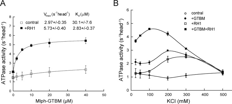
Vertebrate myosin-5a is an ATP-utilizing processive motor associated with the actin network and responsible for the transport and localization of several vesicle cargoes. To transport cargo efficiently and prevent futile ATP hydrolysis, myosin-5a motor function must be tightly regulated. The globular tail domain (GTD) of myosin-5a not only functions as the inhibitory domain but also serves as the binding site for a number of cargo adaptor proteins, including melanophilin (Mlph) and Rab-interacting lysosomal protein-like 2 (RILPL2). In this study, using various biochemical approaches, including ATPase, single-molecule motility, GST pulldown assays, and analytical ultracentrifugation, we demonstrate that the binding of both Mlph and RILPL2 to the GTD of myosin-5a is required for the activation of myosin-5a motor function under physiological ionic conditions. We also found that this activation is regulated by the small GTPase Rab36, a binding partner of RILPL2. In summary, our results indicate that RILPL2 is required for Mlph-mediated activation of Myo5a motor activity under physiological conditions and that Rab36 promotes this activation. We propose that Rab36 stimulates RILPL2 to interact with the myosin-5a GTD; this interaction then induces exposure of the Mlph-binding site in the GTD, enabling Mlph to interact with the GTD and activate myosin-5a motor activity.

Keywords: Rab; Rab36; actin; allosteric regulation; melanophilin; melanosome; molecular motor; myosin; myosin-5a; organelle; small GTPase; vesicle transport.


Conflict of interest statement

The authors declare that they have no conflicts of interest with the contents of this article

Figures

Figure 1.
Figure 1.
RILPL2 specifically interacts with Myo5a-GTD and enhances the interaction between Myo5a and Mlph-GTBM. A, Myo5a-GTD specifically interacts with RILPL2 but not RILP or RILPL1. Shown is GST pulldown of GST-Myo5a-GTD with RILP, RILPL1, and RILPL2. GST-Myo5a-GTD (2 μm) was incubated with 4 μm His-RILP, His-RILPL1, or His-RILPL2 and then pulled down by GSH-Sepharose. Inputs and pulldown samples were analyzed by SDS-PAGE (4%–20%) and visualized with CBB staining. The bands of ∼43 kDa and ∼26 kDa are the degradation of GST-GTD during the assay. B, RILPL2-RH1 enhances binding of Mlph-GTBM with Myo5a. FLAG-tagged full-length Myo5a (0.5 μm) was incubated with 2 μm GST-Mlph-GTBM and/or 2 μm His-RILPL2-RH1 and then pulled down using GSH-Sepharose. Bottom panel, the inputs and the pulldown samples were analyzed by Western blotting with the indicated antibodies. Top panel, the amount of Myo5a pulled down in the presence of RILPL1-RH1 relative to the absence of RILPL2-RH1 (data are the mean ± S.D. of three independent assays). IB, immunoblot.
Figure 2.
Figure 2.
RILPL2-RH1 enhances the activation of Myo5a ATPase activity by Mlph-GTBM. A, activation of Myo5a ATPase activity by Mlph-GTBM in the absence or presence of RILPL2-RH1. The ATPase activity of Myo5a was measured in the presence of 0–40 μm Mlph-GTBM under EGTA conditions (20 mm MOPS-KOH (pH 7.0), 100 mm KCl, 1 mm DTT, 1 mm MgCl2, 0.25 mg/ml BSA, 2.5 mm PEP, 20 units/ml pyruvate kinase, 12 μm CaM, 0.5 mm ATP, 40 μm actin, and 1 mm EGTA) in the absence (open squares) or presence (closed squares) of 5 μm RILPL2-RH1. Vmax and Kd were obtained by a hyperbolic fit and are shown as means ± SDs from three independent assays. B, the effects of ionic strength on the activation of Myo5a ATPase activity by RILPL2-RH1 and Mlph-GTBM. The ATPase activity of Myo5a was determined under EGTA conditions and 25–500 mm KCl in the absence or presence of Mlph-GTBM and/or RILPL2-RH1. Open circles, in the absence of Mlph-GTBM and RILPL2; closed circles, in the presence of 40 μm Mlph-GTBM; open squares, in the presence of 5 μm His-RILPL2-RH1; closed squares, in the presence of 20 μm Mlph-GTBM and 5 μm His-RILPL2-RH1. All data are means ± SDs from three independent assays.
Figure 3.
Figure 3.
RILPL2-RH1 and Mlph-GTBM synergistically activate Myo5a motility activity. A, total internal reflection fluorescence microscopy images showing the movements of Myo5a molecules (green, Cy3B-Myo5a) along actin filaments (red, Alexa 488–labeled F-actin) in the absence or presence of 20 μm Mlph-GTBM and/or 5 μm RILPL2-RH1. The moving spots are indicated by arrowheads (see also Movies S1–S4). For clarity, only a part of the image (150 × 150 pixels) is shown. Scale bars = 5 μm. B, normalized frequency of Cy3B fluorescent spots moving along Alexa 488–labeled F-actin within a fixed time frame and area. Values are normalized to a 1-mm length of actin filament in 1 min. Values are means ± SDs of three different movies.
Figure 4.
Figure 4.
RILPL2-RH1 and Mlph-GTBM synergistically induce Myo5a to form the extended conformation. The S values of Myo5a (∼2 μm Myo5a in 20 mm MOPS (pH 7.0), 100 mm KCl, 1 mm EGTA, 1 mm MgCl2, and 1 mm DTT) in the absence or presence of RILPL2-RH1 and Mlph-GTBM were measured by analytical ultracentrifugation. Control, in the absence of RILPL2-RH1 and Mlph-GTBM; +RH1, in the presence of 10 μm RILPL2-RH1; +GTBM, in the presence of 10 μm Mlph-GTBM; +RH1+GTBM, in the presence of 5 μm RILPL2-RH1 and 5 μm Mlph-GTBM.
Figure 5.
Figure 5.
Rab36 stimulates Mlph-GTBM/RILPL2–mediated activation of Myo5a ATPase activity by enhancing RILPL2's interaction with Myo5a. A, Rab36 interacts with RILPL2 in a GTP-dependent manner. GST or GST-RILPL2 (20 μm, 20 μl) was bound onto 10 μl of GSH-Sepharose and then incubated with 200 μl of 2 μm His-Rab36 preloaded with GTPγS or GDP. The GSH-Sepharose–bound proteins were eluted by GSH and then analyzed by SDS-PAGE and visualized with CBB staining. B, the effects of Rab36 on Mlph-GTBM/RILPL2–mediated activation of Myo5a ATPase activity. The ATPase activity of Myo5a was measured under EGTA conditions as described in Fig. 1A, except that 5 μm His-RILPL2-RH1, 5 μm Mlph-GTBM, 5 μm His-RILPL2, and/or 20 μm Rab36-GTPγS were added. C, Rab36 enhances RILPL2's interaction with Myo5a-GTD. GST-Myo5a-GTD (4 μm) was incubated with 10 μm His-RILPL2 and/or 10 μm His-RILPL2-RH1 and/or 15 μm His-Rab36-GTPγS and then pulled down using GSH-Sepharose. Left panel, the inputs and the pulldown samples were separated by SDS-PAGE and visualized with CBB staining. Right panel, the molar ratio of RILPL2 versus RILPL2-RH1 pulled down with GST-Myo5a-GTD in the presence of 0–15 μm His-Rab36-GTPγS. The amounts of RILPL2 and RILPL2-RH-1 pulled down were quantified using the National Institutes of Health ImageJ program. The molar ratio of RILPL2 versus RILPL2-RH1 was calculated based on their molecular masses. Data are means ± SDs of at least three independent assays.
Figure 6.
Figure 6.
Proposed model for the regulation of Myo5a by RILPL2 and Mlph. A, the structures of the Myo5b-GTD homodimer and Myo5a-GTD/RILPL2-RH1/Mlph-GTBM ternary complex. Top, the structure of the Myo5b-GTD homodimer (PDB code 4J5M) formed by two symmetry-related molecules. Bottom, the structure of the Myo5a-GTD/RILPL2-RH1/Mlph-GTBM ternary complex (PDB code 4KP3). The Mlph-GTBM–binding site and the putative motor domain–binding site are indicated. Sub-1 and Sub-2 indicate subdomain 1 and 2 of Myo5a-GTD, respectively. B, a three-state model for the activation of Myo5a motor function by Rab36, RILPL2, and Mlph-GTBM. In the inhibited state, the two Myo5a-GTDs form a dimer and bind to the two heads of Myo5a, forming a triangular folded conformation. RILPL2 contains two domains, RH1 and RH2, which interact with Myo5a-GTD and the GTP-bound Rab36, respectively. In the absence of Rab36, RH2 prevents RH1 from binding to Myo5a-GTD. Binding of GTP-bound Rab36 to RH2 relieves RH2's inhibition on RH1. RH1 then binds to the GTD dimer of Myo5a in the inhibited state and induces the GTD to expose the Mlph-GTBM–binding site, forming the preactivated state. Finally, Mlph-GTBM binds to the Mlph-GTBM–binding site of Myo5a-GTD in the preactivated state and induces Myo5a to transform to the activated state in the extended conformation.

References

    1. Mehta A. D., Rock R. S., Rief M., Spudich J. A., Mooseker M. S., and Cheney R. E. (1999) Myosin-V is a processive actin-based motor. Nature 400, 590–593 10.1038/23072 - DOI - PubMed
    1. Ikebe M. (2008) Regulation of the function of mammalian myosin and its conformational change. Biochem. Biophys. Res. Commun. 369, 157–164 10.1016/j.bbrc.2008.01.057 - DOI - PubMed
    1. Sellers J. R., Thirumurugan K., Sakamoto T., Hammer J. A. 3rd, Knight P. J. (2008) Calcium and cargoes as regulators of myosin 5a activity. Biochem. Biophys. Res. Commun. 369, 176–181 10.1016/j.bbrc.2007.11.109 - DOI - PubMed
    1. Trybus K. M. (2008) Myosin V from head to tail. Cell Mol. Life Sci. 65, 1378–1389 10.1007/s00018-008-7507-6 - DOI - PMC - PubMed
    1. Zhang N., Yao L. L., and Li X. D. (2018) Regulation of class V myosin. Cell Mol. Life Sci. 75, 261–273 10.1007/s00018-017-2599-5 - DOI - PMC - PubMed

Publication types

MeSH terms