. 2019 Jun 7;10(1):2493.
doi: 10.1038/s41467-019-10355-1.

Tau local structure shields an amyloid-forming motif and controls aggregation propensity

Affiliations

Tau local structure shields an amyloid-forming motif and controls aggregation propensity

Dailu Chen et al. Nat Commun. .

Abstract

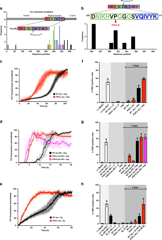
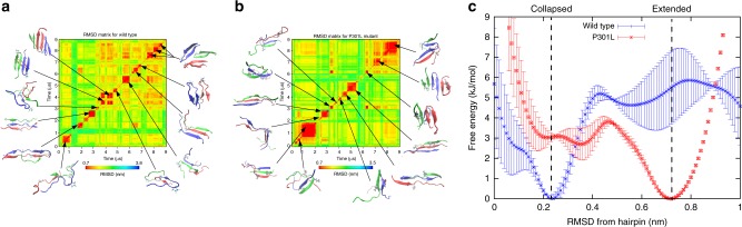
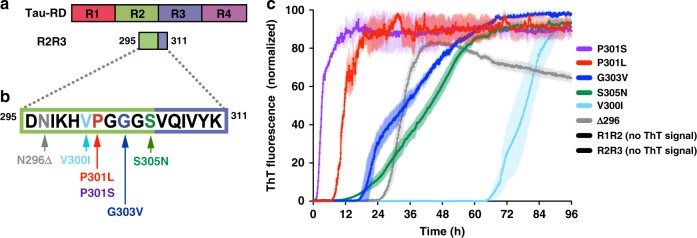
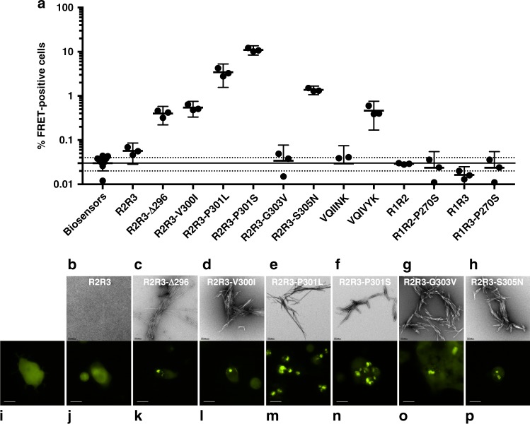
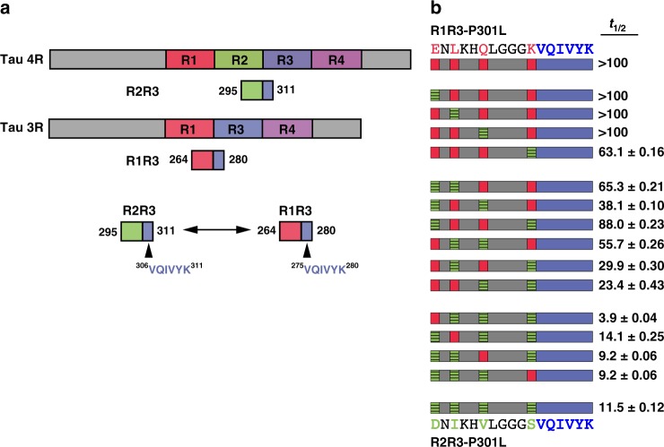
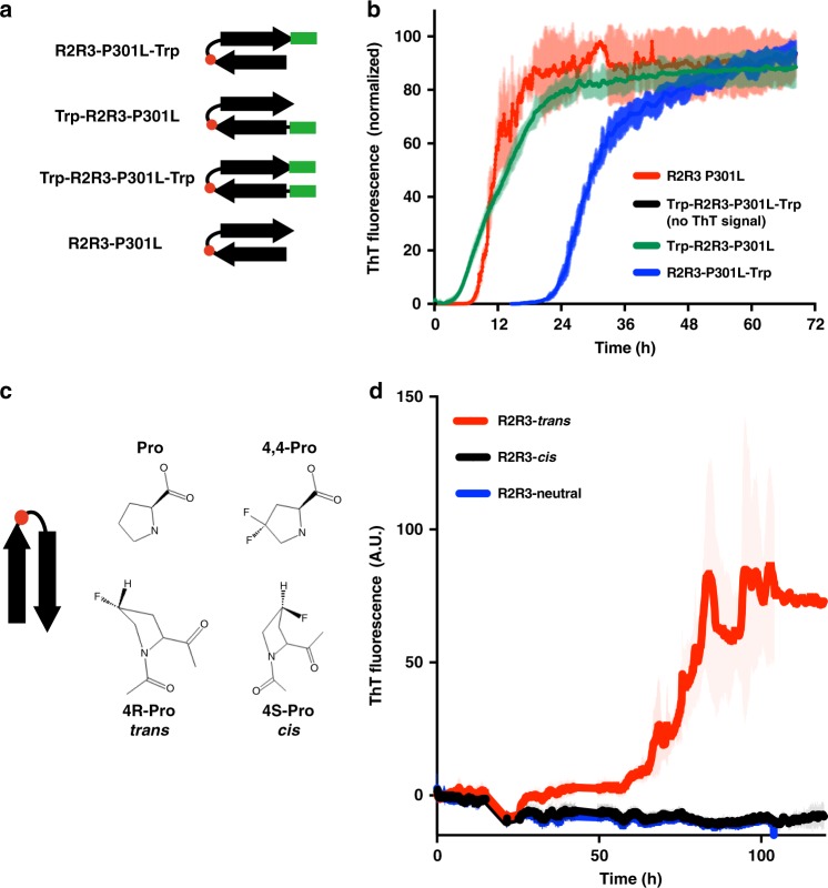
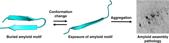
Tauopathies are neurodegenerative diseases characterized by intracellular amyloid deposits of tau protein. Missense mutations in the tau gene (MAPT) correlate with aggregation propensity and cause dominantly inherited tauopathies, but their biophysical mechanism driving amyloid formation is poorly understood. Many disease-associated mutations localize within tau's repeat domain at inter-repeat interfaces proximal to amyloidogenic sequences, such as 306VQIVYK311. We use cross-linking mass spectrometry, recombinant protein and synthetic peptide systems, in silico modeling, and cell models to conclude that the aggregation-prone 306VQIVYK311 motif forms metastable compact structures with its upstream sequence that modulates aggregation propensity. We report that disease-associated mutations, isomerization of a critical proline, or alternative splicing are all sufficient to destabilize this local structure and trigger spontaneous aggregation. These findings provide a biophysical framework to explain the basis of early conformational changes that may underlie genetic and sporadic tau pathogenesis.


Conflict of interest statement

The authors declare no competing interests.

Figures

Fig. 1
Fig. 1
Tauopathy mutations cluster to inter-repeat regions and promote aggregation. a Disease-associated mutation frequency found in human tauopathies. Most mutations are found within the repeat domain (tau RD) (repeat 1 = red; repeat 2 = green; repeat 3 = blue; repeat 4 = purple). Amyloidogenic sequence 306VQIVYK311 is shown in the inset cartoon. b Detailed mutation frequencies found near the 306VQIVYK311 amyloid motif. c FL WT tau and mutant P301L tau at a 4.4 µM concentration were mixed with stoichiometric amounts of heparin (4.4 µM), and allowed to aggregate in the presence of ThT at room temperature. Control WT and P301L tau in the absence of heparin yielded no detectible ThT signal change (less than twofold ratio to background signal) over the course of the experiment (see Supplementary Data 1). ThT fluorescence was normalized to the maximum for each condition. d WT tau RD and mutant P301L and P301S tau RD at a 4.4 µM concentration were each mixed with equimolar amounts of heparin (4.4 µM), and allowed to aggregate in the presence of ThT at room temperature. Control WT, P301L, and P301S tau RD in the absence of heparin yielded no detectible ThT signal change (less than twofold ratio to background signal) over the course of the experiment (see Supplementary Data 1). e WT FL tau and mutant P301L tau at a 4.4 µM concentration were mixed with sub-stoichiometric Ms tau seed (33 nM) and allowed to aggregate in the presence of ThT at room temperature. Control WT and P301L tau in the absence of Ms yielded no detectible ThT signal change (less than twofold ratio to background signal) over the course of the experiment (see Supplementary Data 1). All ThT experiments were carried out in triplicate. The data are shown as the average with standard deviation and are colored according to mutation. fh After 120 h of in vitro incubation, proteins from previous ThT experiments were transduced into tau biosensor cells via lipofectamine (Methods). FRET signal from each condition (tau RD-CFP/tau RD-YFP) was measured by flow cytometry on three biological triplicates of at least 10,000 cells per condition. Error bars represent a 95% CI of each condition
Fig. 2
Fig. 2
Tau RD encodes global and local structure. a Cartoon schematic of tau RD used for XL-MS studies colored according to repeat domain. Recombinant WT and P301L tau RD were heated at 37 °C, 50 °C or 75 °C for 1 hour, then chemically cross-linked using DSS. After cross-linking, trypsin fragmentation, and LC-MS/MS analysis were performed. Each sample was carried out in five technical replicates. b Total consensus cross-links parsed by temperature and location in WT and P301L tau RD: within N-terminus (blue; residues 243–310; N-term), within C-terminus (orange; residues 311–380; C-term), span N- and C-terminus (magenta; between residues 243–310 and 311–380; N-C) and between repeat 2 and repeat 3 (R2R3) (gray; between residues 275–305 and 306–336). ce Consensus cross-links (circles) are shown in contact maps color coded by average frequency across replicates. The theoretical lysine pairs are shown in the background as gray circles. Cross-link contacts within the N-term (blue), C-term (red), and across N- to C-term (purple) are shown as sectors. The x and y axis are colored according to repeat number as in Fig. 1. The dashed boxes define inter-repeat cross-links observed between repeat 2 and repeat 3. fh Same as ce above, except with tau RD that contains a P301L mutation
Fig. 3
Fig. 3
Wild-type and mutant peptides differentially populate collapsed and extended conformations. a Trimer conformations obtained from MD simulations of WT peptide fragment (R2R3-WT) with the sequence 295DNIKHVPGGGSVQIVYK311. Two-dimensional root mean-squared-differences (RMSD’s) are calculated between all pairs of conformations visited during MD simulations. Snapshots of trimeric structures are depicted for selected metastable basins, with each peptide monomer represented by a different color. b The same analysis as in a, but for the P301L substituted trimer. c The free-energy surface as a function of deviation from a canonical hairpin structure. Two distinct basins, corresponding to collapsed and extended sub-ensembles, are found in WT and P301L peptide fragment, respectively. Error bars represent a 95% CI of each RMSD bin
Fig. 4
Fig. 4
Tauopathy mutations drive aggregation propensity. a Schematic of tau RD and the derived peptides representing the minimal structural element around 306VQIVYK311. b WT and mutant peptides were disaggregated, resuspended to 200 µM, and allowed to aggregate in the presence of ThT at room temperature. The WT R2R3 and R1R2 fragment peptides yielded no detectible ThT signal change (less than twofold ratio to background signal) over the course of the experiment (see Supplementary Data 1). ThT signals are shown as average of triplicates with standard deviation, are colored according to mutation and are normalized to the maximum for each condition
Fig. 5
Fig. 5
Peptides form amyloid structures and seed in vivo. a After 96 h of in vitro incubation, peptides from previous ThT experiments (Fig. 4c) were transduced into tau biosensor cells via lipofectamine (Methods). FRET signal from each condition (tau RD-CFP/tau RD-YFP) was measured by flow cytometry on three biological triplicates of at least 10,000 cells per condition. Error bars represent a 95% CI of each condition. Solid and dashed horizontal lines represent the mean and 95% error from untreated biosensor cells, respectively, for ease of statistical comparison. bh Electron microscopy images of each peptide from previous ThT experiments (Fig. 4c). The black bar represents 200 nm distance in each image. ip Qualitative fluorescence microscopy images of tau biosensor cells immediately prior to flow cytometry experiments. i shows a representative image of untreated biosensor cells. jp each shows a representative image of biosensor cells treated with samples from bh, respectively. The white bar represents 10 μm distance in each image
Fig. 6
Fig. 6
Alternative splicing modulates aggregation propensity. a Cartoon schematic for tau 4R and 3R splice isoforms illustrate the difference in primary amino-acid sequence leading into the amyloidogenic 306VQIVYK311 motif. b A full combinatorial panel of R2R3-P301L and R1R3-P301L chimeras were aggregated in vitro. 306VQIVYK311 is shown in blue, amino acids common between the splice isoforms are shown in gray, amino acids unique to an R3 isoform are colored red, amino acids unique to an R4 isoform are colored green. The aggregation kinetics, represented as t1/2 in hours with 95% CI, are listed in the right-side column alongside its respective peptide
Fig. 7
Fig. 7
Enhancing β-hairpin structure rescues spontaneous aggregation phenotypes. a Cartoon schematic representation of the tryptophan zipper motif (green bar) and controls used to stabilize a β-hairpin structure in an R2R3-P301L peptide fragment (Supplementary Table 2). b Aggregation reactions of the tryptophan zipper peptide and controls measured by ThT fluorescence. The Trp-R2R3-P301L-Trp fragment peptide yielded no detectible ThT signal change (less than twofold ratio to background signal) over the course of the experiment (see Supplementary Data 1) ThT signals are shown as average of triplicates with standard deviation and were normalized to the maximum for each condition. c Schematic of proline and fluorinated proline analogs used to generate cis and trans proline conformers at the position corresponding to P301 (red circle) in peptide models. d ThT aggregation reactions of the cis, trans, and neutral proline analogs substituted into the R2R3 peptide fragment. ThT signals are an average of six independent experiments with standard deviation shown
Fig. 8
Fig. 8
Molecular model of tau amyloid domain structural rearrangement and subsequent aggregation. Naive tau monomer (left) exists with a propensity to form a relatively collapsed structure, which buries the amyloid domain 306VQIVYK311. In the presence of disease-associated mutations, proline isomerization events, or certain splice isoforms, the equilibrium is shifted to disfavor local compact structure. This exposes the aggregation-prone 306VQIVYK311 amyloid motif and enhances aggregation propensity, leading to subsequent tau pathology

References

    1. Cleveland DW, Hwo SY, Kirschner MW. Physical and chemical properties of purified tau factor and the role of tau in microtubule assembly. J. Mol. Biol. 1977;116:227–247. doi: 10.1016/0022-2836(77)90214-5. - DOI - PubMed
    1. Eliezer D, et al. Residual structure in the repeat domain of tau: Echoes of microtubule binding and paired helical filament formation. Biochemistry. 2005;44:1026–1036. doi: 10.1021/bi048953n. - DOI - PubMed
    1. Fitzpatrick AWP, et al. Cryo-EM structures of tau filaments from Alzheimer’s disease. Nature. 2017;56:343. - PMC - PubMed
    1. Sanders DW, et al. Distinct Tau prion strains propagate in cells and mice and define different tauopathies. Neuron. 2014;82:1271–1288. doi: 10.1016/j.neuron.2014.04.047. - DOI - PMC - PubMed
    1. Sawaya MR, et al. Atomic structures of amyloid cross-beta spines reveal varied steric zippers. Nature. 2007;447:453–457. doi: 10.1038/nature05695. - DOI - PubMed

Publication types

MeSH terms