

Clinical features of pseudocirrhosis in metastatic breast cancer
- PMID: 31175499
- PMCID: PMC6664810
- DOI: 10.1007/s10549-019-05311-y
Clinical features of pseudocirrhosis in metastatic breast cancer
Abstract
Purpose: Pseudocirrhosis has been demonstrated to mimic cirrhosis radiographically, but studies evaluating the pathophysiology and clinical features are lacking. To better understand the incidence, risk factors, clinical course, and etiology of pseudocirrhosis, we performed a retrospective analysis of consecutively treated patients with metastatic breast cancer (MBC).
Methods: Of 374 patients treated for MBC from 2006 to 2012, 199 had imaging available for review. One radiologist evaluated computed tomography scans for evidence of pseudocirrhosis. Features of groups with and without pseudocirrhosis were compared by Kaplan-Meier product-limit survival estimates and log-rank tests. Wilcoxon Rank-Sum testing evaluated if patients more heavily treated were more likely to develop pseudocirrhosis. Univariate and multivariate Cox proportional hazard models investigated factors associated with mortality.
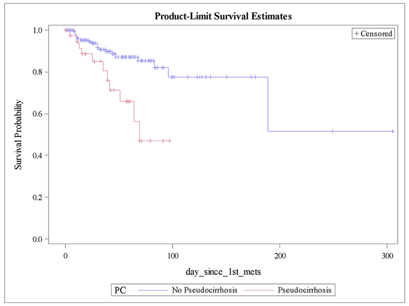
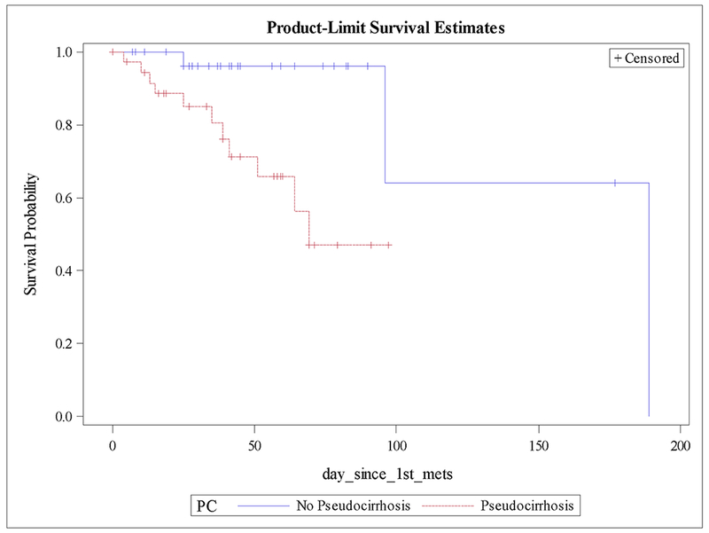
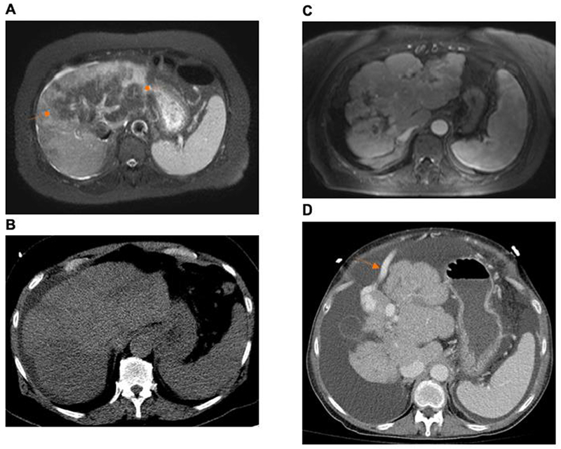
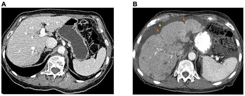
Results: Pseudocirrhosis developed in 37 of 199 patients (19%). Of the patients with liver metastases, 55% developed pseudocirrhosis. Liver metastases were demonstrated in 100% of patients with pseudocirrhosis. Survival in the subset with liver metastases favored those without pseudocirrhosis, 189 versus 69 months (p = 0.01). The number of systemic regimens received were higher in patients with pseudocirrhosis (p = 0.01). Ascites was demonstrated in 68%, portal hypertension in 11%, and splenomegaly in 8% of patients with pseudocirrhosis.
Conclusions: Pseudocirrhosis does not occur in the absence of liver metastases, can manifest as hepatic decompensation, and appears to be associated with poorer survival amongst patients with hepatic metastases. Higher cumulative exposure to systemic therapy may be causative, instead of the previously held belief of pseudocirrhosis as an adverse effect of a particular systemic agent/class.
Keywords: Breast cancer; Cirrhosis; Liver injury; Pseudocirrhosis.
Conflict of interest statement
The authors have no conflicts of interest to report.
Figures
References
-
- Chia SK, Speers CH, D’yachkova Y, et al. The impact of new chemotherapeutic and hormone agents on survival in a population-based cohort of women with metastatic breast cancer. Cancer. 2007. September 1;110(5):973–9. - PubMed
-
- Busni NA: Hepar lobatum carcinomatosum. Virchows Arch A Pathol Anat Histopathol 1924;252:727–733. - PubMed
-
- Symmers D, Spain DM: Hepar lobatum: clinical significance of the anatomic changes. Arch Pathol 1946;42:64–68. - PubMed
-
- Honma K Hepar lobatum carcinomatosum due to metastatic breast carcinoma. Virchows Arch A Pathol Anat Histopathol. 1987;410(6):465–9. - PubMed
