. 2019 Sep;112(3):881-895.
doi: 10.1111/mmi.14324. Epub 2019 Jun 20.

How FtsEX localizes to the Z ring and interacts with FtsA to regulate cell division

Affiliations

How FtsEX localizes to the Z ring and interacts with FtsA to regulate cell division

Shishen Du et al. Mol Microbiol. 2019 Sep.

Abstract

In Escherichia coli, FtsEX, a member of the ABC transporter superfamily, is involved in regulating the assembly and activation of the divisome to couple cell wall synthesis to cell wall hydrolysis at the septum. Genetic studies indicate FtsEX acts on FtsA to begin the recruitment of the downstream division proteins but blocks septal PG synthesis until a signal is received that divisome assembly is complete. However, the details of how FtsEX localizes to the Z ring and how it interacts with FtsA are not clear. Our results show that recruitment of FtsE and FtsX is codependent and suggest that the FtsEX complex is recruited through FtsE interacting with the conserved tail of FtsZ (CCTP), thus adding FtsEX to a growing list of proteins that interacts with the CCTP of FtsZ. Furthermore, we find that the N-terminus of FtsX is not required for FtsEX localization to the Z ring but is required for its functions in cell division indicating that it interacts with FtsA. Taken together, these results suggest that FtsEX first interacts with FtsZ to localize to the Z ring and then interacts with FtsA to promote divisome assembly and regulate septal PG synthesis.


Figures

Fig. 1.
Fig. 1.
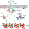
Model for the role of FtsEX in divisome assembly and activation. FtsEX plays three roles in divisome function in E. coli: 1) FtsEX localizes to the Z ring, recruits EnvC and acts on FtsA to promote the monomeric form which is active in recruitment of downstream proteins; 2) FtsEX is involved in transmitting the signal from FtsN to activate FtsWI to synthesize septal PG; and 3) FtsEX regulates amidases (AmiB/A) to hydrolyze peptidoglycan. The latter two roles require the ATPase activity of FtsEX. In the absence of the ATPase activity septal PG synthesis is not initiated and AmiB is not localized.
Fig. 2.
Fig. 2.
The localization of FtsE and FtsX to the Z ring is codependent. Overnight cultures of SD220 (ΔftsEX) carrying plasmids expressing gfp-ftsE (pSD229), gfp-ftsEX (pSD241), ftsX-gfp (pSD226) or ftsEX-gfp (pSD242) were diluted 1:200 in LB with 0.2 M sucrose and ampicillin. After growth at 30 °C for 1 hour, IPTG was added to a final concentration of 15 μM. Two hours post induction, 2 μl of cells were immobilized on an agarose pad containing LB with 0.2 M sucrose to examine the cell morphology and determine the localization of the fusion proteins. Scale bar, 3 μm.
Fig. 3.
Fig. 3.
FtsEX-GFP depends on FtsZ and ZipA for localization. Overnight cultures of W3110, PS106 (ftsZ84ts), PS223 (zipA1ts) and PS236 (ftsA12ts) carrying plasmid pSD242 (pDSW210, P206::ftsEX-gfp) were diluted 100X in fresh LB medium with antibiotics and grown at 30°C for 2 h. The cultures were then diluted 1:10 in fresh LB medium with antibiotics and IPTG was added to a final concentration of 15 μM. Two hours post induction, the cultures were split in half. One half was kept at 30 °C, while the other half was shifted to 42°C. After 30 minutes, cells growing at 30 °C were taken for photography while cells growing at 42 °C were fixed with paraformaldehyde and glutaraldehyde before photographing. Scale bar, 3 μm.
Fig. 4.
Fig. 4.
FtsEX-GFP disrupts Z rings assembled with only FtsA (ZipAts strain). Overnight cultures of SD335 (zapA-mCherry), SD336 (ftsZ84 [Ts], zapA-mCherry), SD337 (ftsA12 [Ts], zapA-mCherry) and SD338 (zipA [Ts], zapA-mCherry) carrying plasmid pSD242 (pDSW210, P206::ftsEX-gfp) were diluted 1:100 in fresh LB medium with antibiotics and grown at 30°C for 2 hours. The cultures were then diluted 1:10 in fresh LB medium with antibiotics and IPTG was added to a final concentration of 15 μM. The cultures were split in half 2 hours later and one half was kept at 30°C, while the other half was shifted to 42°C. Thirty minutes later cells growing at 30°C were taken for photography while cells growing at 42°C were fixed with paraformaldehyde and glutaraldehyde before photographing. Samples from the 30°C cultures are shown in Fig. S3. Scale bar, 3 μm.
Fig. 5.
Fig. 5.
Moderate overexpression of ftsEX-gfp blocks cell division without disrupting Z rings. (A) The sensitivity of strains to expression of ftsEX-GFP was assessed at permissive temperature with a spot assay. In this and all subsequent spot assays cultures were serially diluted 10-fold in LB and spotted on LB plates with antibiotics with and without IPTG. The strains tested were SD335 (zapA-mcherry), PS106 (ftsZ84 [Ts], zapA-mcherry), SD337 (ftsA12 [Ts], zapA-mcherry) and SD338 (zipA1 [Ts], zapA-mcherry) carrying plasmid pDSW210 (P206::gfp) or pSD242 (P206::ftsEX-gfp). Plates were incubated at 30°C overnight before photographing. (B) The effect of ftsEX-gfp expression on Z rings. The strains in (A) were grown to exponential phase in LB with ampicillin and 60 μM IPTG was added. Two hours later samples were taken and cells examined by phase and fluorescence microscopy. Results with strain SD335, SD337 and SD338 are shown. Scale bar, 5 μm
Fig. 6.
Fig. 6.
Moderate overexpression of ftsEX-gfp competes with FtsA at the Z ring. Strains S3 (WT) and PS236 (ftsA12 [Ts]) containing a plasmid expressing ftsEX-gfp (pSD242) and growing exponentially at 30 °C in LB with ampicillin were induced with 60 μM IPTG for 2 hours. Cells were fixed with glutaraldehyde and paraformaldehyde and immunostained for FtsZ, ZipA and FtsA and examined by phase and fluorescence microscopy.
Fig. 7.
Fig. 7.
Higher overexpression of ftsEX blocks cell division by preventing Z ring formation. (A) Spot test to determine the toxicity of ftsE, ftsX and ftsEX overexpression. Cultures of strain W3110 containing plasmids expressing ftsE (pSEB426[P204::ftsE]), ftsX (pSEB427[P204::ftsX]), ftsA (pSEB306+[P204::ftsA]), ftsEX (pSEB428 [pDSW208, P204::ftsEX]) or gfp (pDSW208, P204::gfp) growing in LB with ampicillin were induced with IPTG. Plasmids expressing gfp or ftsA were used as controls. (B) Samples from liquid cultures of some of the strains in (A) were taken two hours after the addition of IPTG (1000 μM), immunostained for FtsZ and analyzed by phase and fluorescence microscopy. A control sample of W3110 treated with 20 μg/ml of cephalexin for 2 hours was also analyzed as a control.
Fig. 8.
Fig. 8.
Model of FtsEX and identification of a region in FtsE that is important for localization of GFP-FtsEX to the Z ring. (A) Model of FtsEX. A model of FtsE was generated by using the nucleotide component of an ABC transporter from Aquifex aeolicus (2PCL) and then superimposing it on an ABC transporter complex from Staphylococcus aureus (PDB:2ONJ). A model of the FtsX monomer was generated using I-TASSER and it along with the FtsE dimer model were superimposed on the structure of MacB (Crow et al., 2017). FtsE is in pink and FtsX is colored cyan. The first 46 residues of FtsX are not in the model and are indicated by a dotted line. The residues important for localization of FtsEX to the Z ring are shown in space filled (magenta and light orange). The region in FtsX colored red is required for the interaction between FtsX and EnvC (residues 152–161)(Yang et al., 2011). (B) Effect of ftsE mutations on the localization of GFP-FtsEX. SD220 (W3110 leu::Tn10, ftsEX::cat) containing pSD241 or derivatives with ftsE mutations were grown in LB with ampicillin and 0.2 M sucrose. Localization was determined following low level of expression (15 μM IPTG) from pSD241 (P206::gfp-ftsEX) and its derivatives. The altered residues are indicated in light orange in panel A. Scale bar, 3 μm.
Fig. 9.
Fig. 9.
The effect of ftsE mutations on complementation, toxicity of FtsED162NX and the interaction of FtsE with FtsZ. (A) Effect of ftsE mutations on complementation. The ability to complement ΔftsEX was assessed in strain SD220 (W3110 leu::Tn10, ftsEX::cat) using plasmids pSD221 (pEXT22, Ptac::ftsEX), expressing ftsEX or a derivative pSD221-EF/KE expressing ftsEE73K,F76EX. Single colonies were picked into LB, diluted in 10 fold increments and spotted on LB plates containing ampicillin with and without sucrose. Plates were incubated at 37 °C overnight before photographing. (B) The effect of ftsE mutations on the toxicity of the ATPase mutant of FtsEX. Plasmid pSD221 or derivatives expressing various alleles of ftsEX were transformed into strain S3 and the toxicity assessed with a spot assay. The plates were incubated at 37 °C overnight before photographing. (C) The effect of various ftsE mutations on the interaction between FtsE (interaction is only observed if ftsE is expressed with ftsX) and FtsZ was assessed with the BACTH. Plasmid pairs were co-transformed into BTH101. The next day, single colonies of each strain were resuspended in 1 ml LB medium and 3 μl of each aliquot was spotted on LB plates containing ampicillin, kanamycin, 40 μg/ml X-gal and 100 μM IPTG. Plates were incubated at 30°C overnight before imaging. (D) Quantification of the interactions in panel C was determined by a β-galatosidase assay as described in Experimental Procedures.
Fig. 10.
Fig. 10.
The N-terminal cytoplasmic domain of FtsX (FtsXNcyto) is dispensable for FtsEX localization but is required for FtsEX function. (A) Complementation test of ftsEXΔ4–69. Plasmids expressing ftsEX (pSD221[Ptac::ftsEX]) or ftsEXΔ4−69 (pSD318[Ptac::ftsEXΔ4−69]) were transformed into strain SD220 (W3110 leu::Tn10, ftsEX::cat). Single transformants were analyzed by a spot assay on LB plates containing kanamycin with or without sucrose. (B) Localization of FtsEXΔ4−69-GFP in ΔftsEX cells. Overnight cultures of SD220 carrying plasmids pSD226 (P206::ftsX-gfp), pSD242 (P206::ftsEX-gfp) or pSD321(P206::ftsEXΔ4−69-gfp) were diluted 1:200 in LB with 0.2 M sucrose and ampicillin. After growth at 30 °C for 1 h, 15 μM IPTG was added to the culture and two hours later cells were analyzed by phase and fluorescence microscopy. (C) FtsXNcyto is required for recruitment of downstream division proteins. Overnight cultures of SD446 (W3110, zapA-mcherry cat<>frt, ftsEX<>frt Para::ftsEX-cat) carrying plasmids pSD332 (pSC101, Psyn:gfp-ftsB) and pEXT22 or derivatives expressing alleles of ftsEX (pSD221 or pSD318) were diluted 1:100 in LB medium with antibiotics and 0.2% arabinose and grown at 30°C for 2 hours. Cells were centrifuged, washed 3 times in LB and resuspended in LB with antibiotics and with or without arabinose. After growth at 30°C for 3 hours, samples were taken and analyzed by phase and fluorescence microscopy. Shown are the samples without arabinose, the control with arabinose is shown in Fig. S14.
Fig. 11.
Fig. 11.
The toxicity of the ATPase mutant requires FtsXNcyto. (A) The toxicity of different ftsEX alleles. Derivates of pEXT22 expressing various alleles of ftsX and ftsE were transformed into strain SD221 (W3110, leu::Tn10 ftsA*, ftsEX::cat) and transformants tested by a spot assay. Single colonies of each strain growing on LB with 0.2 M sucrose and kanamycin were resuspended in LB, serially diluted and spotted on plates containing sucrose, kanamycin and increasing concentrations of IPTG. (B) Overnight cultures of strains from (A) were diluted 1:200 in LB with 0.2 M sucrose and kanamycin. After growth at 30 °C for 1 hour, IPTG was added to final concentration of 60 μM. Two hours after the addition of IPTG, samples were analyzed by phase microscopy.
Fig. 12.
Fig. 12.
Model for FtsEX interactions with other division proteins in E. coli. An FtsZ filament is tethered to the membrane by the CCTP of FtsZ interacting with FtsA and ZipA. The CCTP is connected to the tubulin domain of FtsZ by a 50 amino acid intrinsically disordered linker (green dotted line). 2) FtsEX is recruited to the filament through FtsE bound to FtsX interacting with the CCTP. 3) The N-terminal region of FtsX (indicated by the boxed N) interacts with FtsA (region around residue G366) making FtsA less oligomeric. 4) Less oligomeric FtsA recruits the remaining divisome proteins (FtsK, FtsQLB, FtsWI and FtsN).

References

    1. Aarsman ME, Piette A, Fraipont C, Vinkenvleugel TM, Nguyen-Disteche M & den Blaauwen T, (2005) Maturation of the Escherichia coli divisome occurs in two steps. Molecular microbiology 55: 1631–1645. - PubMed
    1. Addinall SG, Bi E & Lutkenhaus J, (1996) FtsZ ring formation in fts mutants. Journal of bacteriology 178: 3877–3884. - PMC - PubMed
    1. Addinall SG, Cao C & Lutkenhaus J, (1997) FtsN, a late recruit to the septum in Escherichia coli. Molecular microbiology 25: 303–309. - PubMed
    1. Addinall SG & Lutkenhaus J, (1996) FtsA is localized to the septum in an FtsZ-dependent manner. Journal of bacteriology 178: 7167–7172. - PMC - PubMed
    1. Arends SJ, Kustusch RJ & Weiss DS, (2009) ATP-binding site lesions in FtsE impair cell division. Journal of bacteriology 191: 3772–3784. - PMC - PubMed

Publication types

MeSH terms

LinkOut - more resources