

Detection of Cells Translocated with Yersinia Yops in Infected Tissues Using β-Lactamase Fusions
- PMID: 31177435
- PMCID: PMC6733027
- DOI: 10.1007/978-1-4939-9541-7_9
Detection of Cells Translocated with Yersinia Yops in Infected Tissues Using β-Lactamase Fusions
Abstract
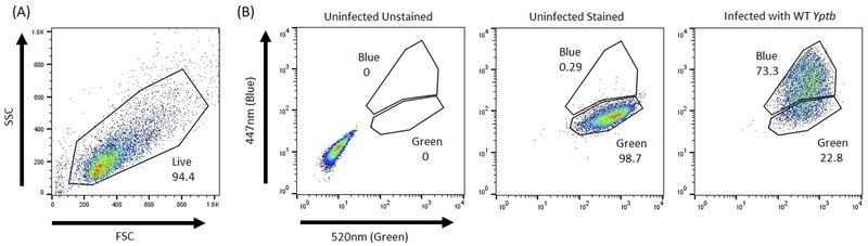
Development of the TEM-CCF2/4-AM FRET-based system has enabled investigators to track translocation of effector proteins into mammalian cells during infection. This allows for separation of translocated and non-translocated cell populations for further study. Yersinia strains expressing translational Yop-TEM fusions, containing the secretion and translocation signals of a Yop with the TEM-1 portion of β-lactamase, are used to infect mice, tissues isolated from mice, or mammalian cells in culture. Infected and harvested mammalian cells are treated with either CCF2-AM or CCF4-AM, and cleavage of this fluorescent compound by TEM is detected by fluorescence-activated cell sorting (FACS) analysis. A shift from green to blue emission spectra of individual cells is indicative of translocation of a given Yop-TEM fusion protein into the host cell during Yersinia infection due to a disruption in FRET between the two fluors of the compound. In Yersinia, this method has been used to understand Type III secretion dynamics and Yop functions in cells translocated by effectors during infection. Here, we describe how to generate Yop-TEM constructs, and how to detect, quantify, isolate, and study Yop-TEM containing cells in murine tissues during infection and in ex vivo tissues by cell sorting and flow cytometry analysis. In addition, we provide guidance for analyzing TEM-positive cells via a plate reader and fluorescent microscopy.
Keywords: Bla; CCF2; CCF4; FACS; Flow cytometry; Neutrophils; T3SS; TEM; Translocation; Type III secretion; Yersinia; Yop translational fusions; Yops; β-Lactamase.
Figures
References
Publication types
MeSH terms
Substances
Grants and funding
LinkOut - more resources
Full Text Sources
Miscellaneous
