. 2019 Aug 13;140(7):566-579.
doi: 10.1161/CIRCULATIONAHA.118.038924. Epub 2019 Jun 10.

Spliced X-box Binding Protein 1 Stimulates Adaptive Growth Through Activation of mTOR

Affiliations

Spliced X-box Binding Protein 1 Stimulates Adaptive Growth Through Activation of mTOR

Xiaoding Wang et al. Circulation. .

Abstract

Background: The unfolded protein response plays versatile roles in physiology and pathophysiology. Its connection to cell growth, however, remains elusive. Here, we sought to define the role of unfolded protein response in the regulation of cardiomyocyte growth in the heart.

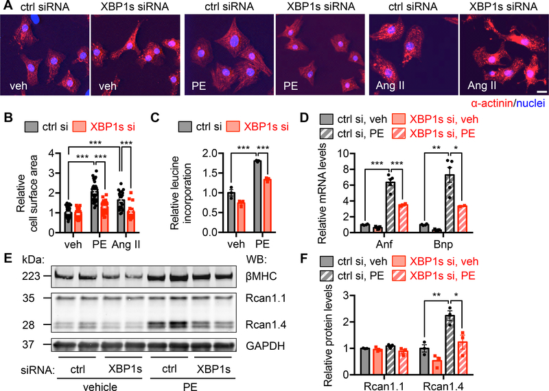
Methods: We used both gain- and loss-of-function approaches to genetically manipulate XBP1s (spliced X-box binding protein 1), the most conserved signaling branch of the unfolded protein response, in the heart. In addition, primary cardiomyocyte culture was used to address the role of XBP1s in cell growth in a cell-autonomous manner.

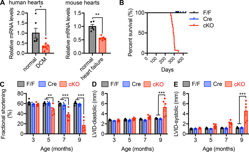
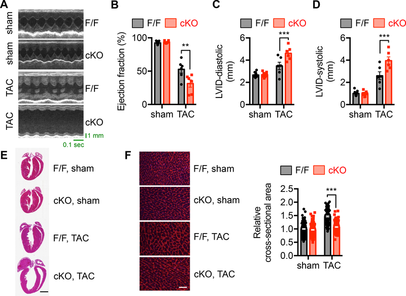
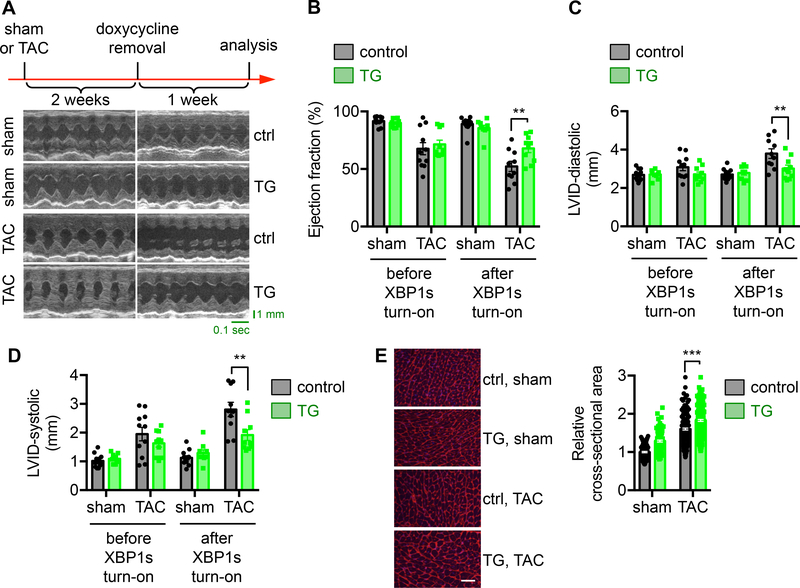
Results: We found that XBP1s expression is reduced in both human and rodent cardiac tissues under heart failure. Furthermore, deficiency of XBP1s leads to decompensation and exacerbation of heart failure progression under pressure overload. On the other hand, cardiac-restricted overexpression of XBP1s prevents the development of cardiac dysfunction. Mechanistically, we found that XBP1s stimulates adaptive cardiac growth through activation of the mechanistic target of rapamycin signaling, which is mediated via FKBP11 (FK506-binding protein 11), a novel transcriptional target of XBP1s. Moreover, silencing of FKBP11 significantly diminishes XBP1s-induced mechanistic target of rapamycin activation and adaptive cell growth.

Conclusions: Our results reveal a critical role of the XBP1s-FKBP11-mechanistic target of rapamycin axis in coupling of the unfolded protein response and cardiac cell growth regulation.

Keywords: FKBP11 protein, human; FKBP11 protein, mouse; X-box binding protein 1; heart failure; hypertrophy; mTOR protein, mouse; unfolded protein response.


Figures

Figure 1.
Figure 1.. Cardiac-specific deficiency of XBP1s leads to cardiomyopathy in mice.
A. XBP1s expression was reduced in the hearts from human patients with dilated cardiomyopathy (DCM). N = 6 for normal controls; 7 for DCM. XBP1s expression was decreased in failing mouse hearts. Cardiac dilation and heart failure were triggered by thoracic aortic constriction (TAC). N = 5 for each group. Student’s t test was conducted. B. Cardiac-specific knockout of XBP1s caused early mortality. XBP1s deletion was achieved by crossing XBP1F/F with the αMHC-Cre mouse models. All conditional knockout (cKO) mice died before 350 days old, while the controls did not show mortality. N = 14 for F/F; 6 for αMHC-Cre; 14 for cKO. The log rank test was conducted. P<0.001 between F/F and cKO, and Cre and cKO, respectively. C. XBP1s cKO mice showed gradual deterioration of cardiac function. Fractional shortening (%) was determined by echocardiography with conscious animals. N = 5 per group. D. XBP1s deficient mice displayed an increase in left ventricular inner diameter (LVID) at diastole. N = 5 for each group. E. XBP1s cKO showed elevated left ventricular diameter at systole. N = 5 for each group. Two-way ANOVA was conducted with the selection of repeated measures, followed by Bonferroni’s multiple comparisons test. *, P<0.05; **, P<0.01; ***, P<0.001.
Figure 2.
Figure 2.. XBP1s deficiency exacerbates heart failure progression by pressure overload.
A. Control and cKO animals of 8 weeks old were used for sham or TAC surgery. Cardiac function was determined 2 weeks later. Representative echocardiography images of control and cKO animals after sham or TAC surgery are shown. B. XBP1s deficiency in the heart led to deterioration of cardiac function and accelerated progression to cardiomyopathy, as revealed by a decrease in ejection fraction (%). N = 6–9. C. Diastolic LVID was significantly elevated in the cKO mice after TAC. N = 6–13. D. LVID at systole was increased by XBP1s deficiency in the heart. N = 6–13. E. Representative cardiac images showed deficiency of ventricular growth in the cKO mice after TAC. Scale bar: 2 mm. F. Wheat germ agglutinin (WGA) staining was performed to visualize cardiac myocytes (left). Scale bar: 50 μm. Quantification is shown at the right. N = 209 for sham/FF; 188 for sham/cKO; 115 for TAC/FF; 143 for TAC/cKO. Two-way ANOVA analysis was conducted, followed by Tukey’s test. **, P<0.01; ***, P<0.001.
Figure 3.
Figure 3.. Overexpression of XBP1s in the heart improves cardiac function in response to pressure overload.
A. XBP1s expression was turned on after 2 weeks of TAC, before the onset of cardiac dysfunction (indicated as doxycycline removal). After another week, control animals (single TRE-XBP1s or αMHC-tTA transgenics alone under water without doxycycline) displayed cardiomyopathy and cardiac dysfunction, while transgenic mice (TRE-XBP1s and αMHC-tTA double transgenics under water without doxycycline) showed significant improvements in cardiac function, as evidenced by representative echocardiographic images. B. Ejection fraction (%) was significantly increased by XBP1s overexpression. N = 9–13. C. LVID at diastole was reduced in the XBP1s transgenic mice after TAC. N = 9–13. D. Systolic LVID in the transgenic mice was improved. N = 9–13. E. WGA staining was conducted (left). Scale bar: 50 μm. Quantification of the relative cross-sectional area shows significant upregulation of cardiomyocyte size by XBP1s overexpression (right). N = 128 for sham/control; 104 for sham/TG; 198 for TAC/control; 226 for TAC/TG. Two-way ANOVA analysis was conducted, followed by Tukey’s test. **, P<0.01; ***, P<0.001.
Figure 4.
Figure 4.. XBP1s is required for cardiac myocyte growth in response to hypertrophic stimuli.
A. Immunofluorescence staining for α-actinin. XBP1s was silenced by siRNA transfection and the cells were treated by phenylephrine (PE, 50 μM) or Angiotensin II (Ang II, 1 μM) for 48 hrs. Scale bar: 20 μm. B. Cardiomyocyte surface area quantification of A. PE or Ang II treatment increased cardiomyocyte size, which was blunted by XBP1s silencing. N = 56 for veh/ctrl si; 57 for veh/XBP1s si; 49 for PE/ctrl si; 50 for PE/XBP1s si; 36 for Ang II/ctrl si; 28 for Ang II/XBP1s si. C. XBP1s knockdown attenuated PE-induced growth as revealed by a 3H-leucine incorporation assay. N = 3 per group. D. Induction of hypertrophic markers by PE was diminished by XBP1s knockdown, as assessed by quantitative RT-PCR. N = 4 for ctrl si; 5 for XBP1s si. E. Immunoblotting showed that the induction of Rcan1.4 by PE was blunted by XBP1s silencing. In contrast, Rcan1.1 expression was not affected. GAPDH was used as a loading control. F. Quantification of E. N = 3 for each group. Two-way ANOVA analysis was performed, followed by Tukey’s test. *, P<0.05; **, P<0.01; ***, P<0.001.
Figure 5.
Figure 5.. XBP1s expression is sufficient to stimulate cardiomyocyte hypertrophic growth.
A. XBP1s overexpression in cardiomyocytes led to an increase in cell size. NRVMs were infected by adenovirus expressing control GFP or XBP1s, followed by PE treatment. Note that the XBP1s-expressing virus is bi-cistronic for GFP and XBP1s, and GFP positivity indicates infection by either GFP-only control or XBP1s-GFP virus. Scale bar: 20 μm. B. Quantification of A. N = 34 for veh/Ad GFP; 33 for veh/Ad XBP1s; 51 for PE/Ad GFP; 107 for PE/Ad XBP1s. C. Protein synthesis was elevated by XBP1s overexpression as revealed by 3H-leucine incorporation. N = 3 for PE/Ad XBP1s. N = 6 for the other groups. D. XBP1s overexpression potentiated PE-induced upregulation of Rcan1.4 at the protein level. As a control, Rcan1.1 did not show a similar trend. E. Quantification of D. N = 5 for Rcan1.4/Ad XBP1s. N = 6 for the other groups. Two-way ANOVA analysis was conducted, followed by Tukey’s test. *, P<0.05; **, P<0.01; ***, P<0.001.
Figure 6.
Figure 6.. XBP1s stimulates mTOR signaling in the heart.
A. Cardiomyocyte-specific overexpression of XBP1s increased mTOR signaling in the heart. XBP1s expression in the heart was turned on for 2 weeks by removal of doxycycline from drinking water. Cardiac tissues were subjected to Western blotting to detect the mTOR signaling. GAPDH was used as loading control. B. Quantification of A. Normalization was done with phosphorylation levels to respective total proteins. Student’s t test was conducted. N = 4–7. C. XBP1s overexpression in vitro augmented mTOR activity. NRVMs were infected by adenovirus expressing either GFP or XBP1s. PE treatment was then conducted for 24 hrs. D. Quantification of C. N = 5 for each group. E. XBP1s knockdown decreased the mTOR activity. XBP1s expression was reduced by siRNA transfection in NRVMs and PE was incubated for 24 hrs. GAPDH was used as a loading control. F. Quantification of E. N = 3–6. Two-way ANOVA analysis was conducted, followed by Tukey’s test. *, P<0.05; **, P<0.01; ***, P<0.001.
Figure 7.
Figure 7.. FKBP11 is a direct target of XBP1s.
A. FKBP11 expression was acutely induced by pressure overload in the heart. TAC was conducted in wild type animals. Hearts were harvested at different time points post surgery. Relative FKBP11 mRNA level was determined by quantitative RT-PCR. Note that XBP1s expression was also upregulated by TAC. Two-way ANOVA analysis was conducted, followed by Tukey’s test. N = 3–8. B. FKBP11 protein level was augmented by TAC in the heart as shown by immunoblotting, along with induction of XBP1s. GAPDH and Histone H3 were used as loading controls for whole cell lysates and nuclear extracts, respectively. C. A conserved region in the second intron of FKBP11 genome, which resembles the consensus sequence of XBP1s-binding site UPRE (unfolded protein response element). The translational initiation site is in the first exon of FKBP11 genomic DNA. D. XBP1s expression stimulated FKBP11 promoter activity in a dose-dependent manner. The conserved region of FKBP11 promoter was engineered into a luciferase reporter construct. Co-transfection of a XBP1s-expressing plasmid led to an increase in luciferase activity. Note that a truncated mutant of FKBP11 promoter lacking the UPRE region did not show significant promoter activity. N = 4 per group. Two-way ANOVA analysis was performed, followed by Tukey’s test. E. Chromatin immunoprecipitation (ChIP) assay was conducted to determine the binding of XBP1s to the FKBP11 promoter. NRVMs were infected by adenovirus expressing flag-tagged XBP1s. ChIP was performed with either control mouse IgG or anti-flag antibody. PCR was conducted using primers spanning the UPRE site. Primers from the distal region were used as a negative control. F. The mRNA level of FKBP11 was upregulated in the XBP1s transgenic hearts, as determined by quantitative RT-PCR. Student’s t test was performed. N = 3–4. G. The protein level of FKBP11 was significantly increased in the XBP1s transgenic hearts, as revealed by immunoblotting. GAPDH was used as a loading control. N = 3 for each group. Student’s t test was conducted. **, P<0.01; ***, P<0.001.
Figure. 8.
Figure. 8.. FKBP11 is required for XBP1s-mediated mTOR activation.
A. FKBP11 was required for XBP1s-induced hypertrophic growth. NRVMs were infected by adenovirus expressing either GFP or XBP1s. FKBP11 was silenced by siRNA transfection. A 3H-leucine incorporation assay was conducted to assess cell growth. N = 3 for each group. B. FKBP11 knockdown led to a decrease in XBP1s-induced mTOR activation. NRVMs were first infected by adenovirus to overexpression control GFP or XBP1s. FKBP11 siRNA was then used to transfect NRVMs and cell lysates were extracted for immunoblotting. GAPDH was used as a loading control. C. Induction of mTOR activity by XBP1s was significantly inhibited by FKBP11 silencing as shown by quantification of B. N = 3–6. D. Silencing of FKBP11 led to a decrease in PE and XBP1s-induced mTOR activation. NRVMs were first infected by adenovirus expressing GFP or XBP1s. FKBP11-specific siRNA was used to transfect the cells. PE treatment was conducted for 24 hrs and immunoblotting was performed to examine the mTOR signaling. E. Quantification of D. N = 3–6. F. Overexpression of XBP1s in NRVMs increased colocalization of mTOR and LAMP2, a lysosomal marker. FKBP11 knockdown significantly diminished this effect. Scale bar: 20 μm. Two-way ANOVA analysis was performed, followed by Tukey’s test. *, P<0.05; **, P<0.01; ***, P<0.001.

References

    1. Benjamin EJ, Virani SS, Callaway CW, Chamberlain AM, Chang AR, Cheng S, Chiuve SE, Cushman M, Delling FN, Deo R, de Ferranti SD, Ferguson JF, Fornage M, Gillespie C, Isasi CR, Jimenez MC, Jordan LC, Judd SE, Lackland D, Lichtman JH, Lisabeth L, Liu S, Longenecker CT, Lutsey PL, Mackey JS, Matchar DB, Matsushita K, Mussolino ME, Nasir K, O’Flaherty M, Palaniappan LP, Pandey A, Pandey DK, Reeves MJ, Ritchey MD, Rodriguez CJ, Roth GA, Rosamond WD, Sampson UKA, Satou GM, Shah SH, Spartano NL, Tirschwell DL, Tsao CW, Voeks JH, Willey JZ, Wilkins JT, Wu JH, Alger HM, Wong SS and Muntner P. Heart Disease and Stroke Statistics-2018 Update: A Report From the American Heart Association. Circulation. 2018;137:e67–e492. - PubMed
    1. Drazner MH. The progression of hypertensive heart disease. Circulation. 2011;123:327–334. - PubMed
    1. Uhlen M, Fagerberg L, Hallstrom BM, Lindskog C, Oksvold P, Mardinoglu A, Sivertsson A, Kampf C, Sjostedt E, Asplund A, Olsson I, Edlund K, Lundberg E, Navani S, Szigyarto CA, Odeberg J, Djureinovic D, Takanen JO, Hober S, Alm T, Edqvist PH, Berling H, Tegel H, Mulder J, Rockberg J, Nilsson P, Schwenk JM, Hamsten M, von Feilitzen K, Forsberg M, Persson L, Johansson F, Zwahlen M, von Heijne G, Nielsen J and Ponten F. Proteomics. Tissue-based map of the human proteome. Science. 2015;347:1260419. - PubMed
    1. Walter P and Ron D. The unfolded protein response: from stress pathway to homeostatic regulation. Science. 2011;334:1081–1086. - PubMed
    1. Hetz C and Mollereau B. Disturbance of endoplasmic reticulum proteostasis in neurodegenerative diseases. Nat Rev Neurosci. 2014;15:233–249. - PubMed

Publication types

MeSH terms