. 2019 Jun 10;7(1):94.
doi: 10.1186/s40478-019-0747-x.

Neutrophil extracellular trap induced by HMGB1 exacerbates damages in the ischemic brain

Affiliations

Neutrophil extracellular trap induced by HMGB1 exacerbates damages in the ischemic brain

Seung-Woo Kim et al. Acta Neuropathol Commun. .

Erratum in

Abstract

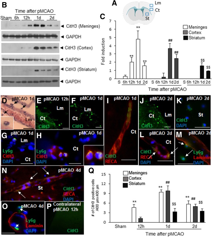
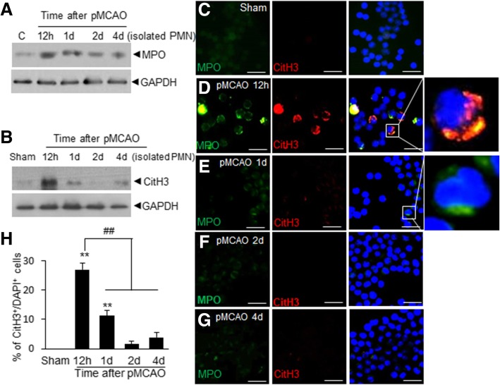
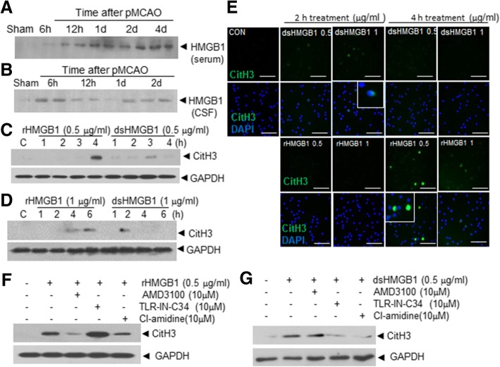
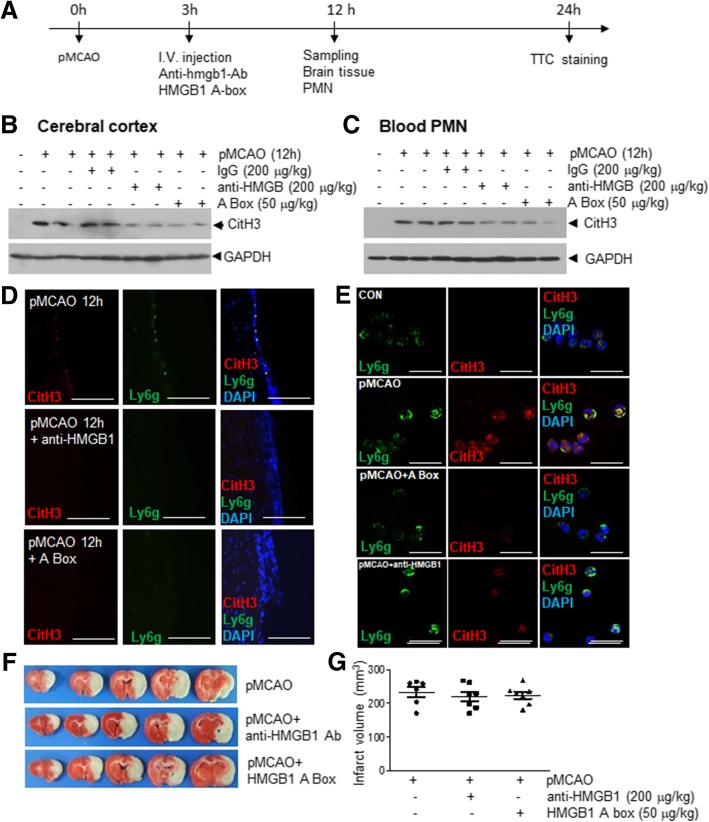
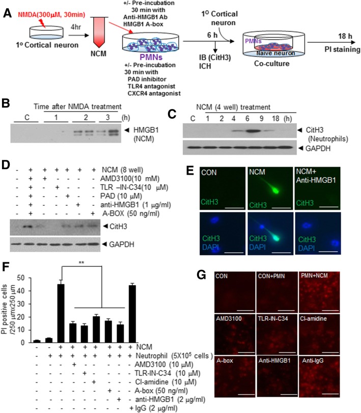
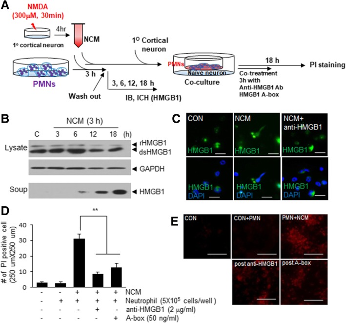
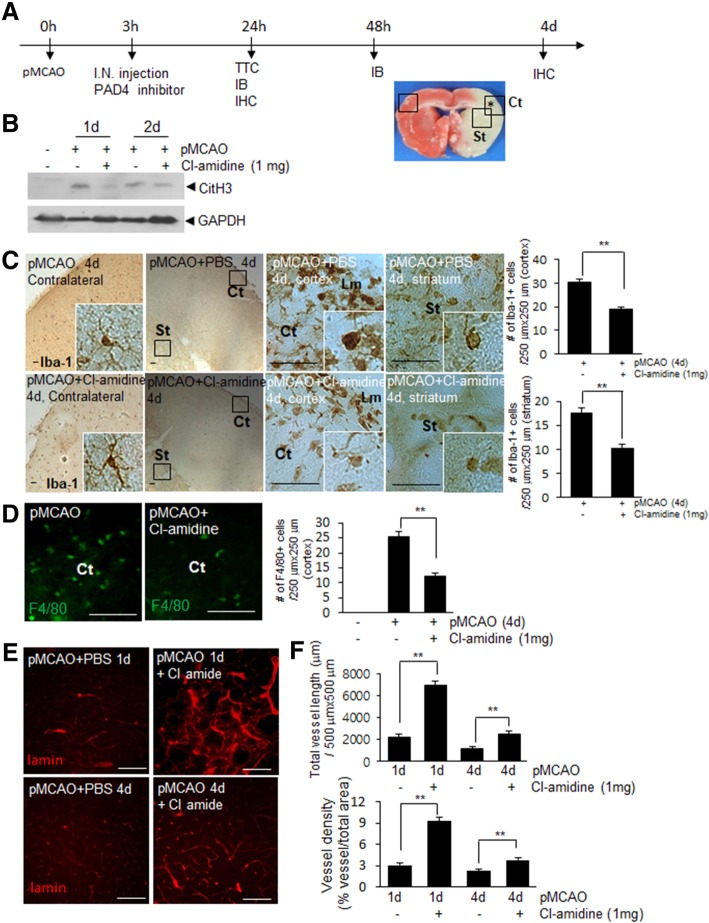
It has been reported that neutrophil extracellular traps (NETs) play important roles in non-infectious diseases. In ischemic stroke, neutrophils infiltrate damaged brain tissue soon after injury and aggravate inflammation. Using a rat permanent MCAO model, we showed citrullinated histone H3+ (CitH3, a marker of NETosis) induction in neutrophils in leptomeninges and in peripheral blood soon after MCAO. Entry of CitH3+ cells occurred through leptomeninges after 6 h of MCAO and these cells were observed in cerebral cortex from 12 h and subsequently in striatum. It is interesting to note that CitH3+ induction began in circulating neutrophils before they migrated to brain parenchyma and they were detected as intact or lysed form. High mobility group box 1 (HMGB1), a danger associated molecular pattern (DAMP) molecule, was accumulated massively in serum after permanent MCAO and plays a critical role in CitH3 inductions in neutrophils in brain parenchyma and in peripheral blood. Both the all-thiol and disulfide types of HMGB1 induced CitH3 via their specific receptors, CXCR4 and TLR4, respectively. Importantly, HMGB1 not only induced NETosis but was included as a part of the extruded NETs, and contribute to NETosis-mediated neuronal death. Therefore, it would appear a vicious cycle exists between neuronal cell death and NETosis and HMGB1 mediates detrimental effects exerted by this cycle. When NETosis was suppressed by a PAD inhibitor in MCAO animals, delayed immune cell infiltrations were markedly suppressed and damages in blood vessels were significantly mitigated. The study shows NETosis with the involvement of HMGB1 as a mediator in a vicious cycle aggravates inflammation and subsequent damage in the ischemic brain.

Keywords: HMGB1; Inflammation; MCAO; NETosis; Permanent ischemia.


Conflict of interest statement

The authors declare that they have no competing interests.

Figures

Fig. 1
Fig. 1
Elevation of CitH3 levels in ischemic brains after MCAO. a Three brain regions used in the present study are indicated as follows, leptomeninges (Lm), cortex (Ct), and striatum (St). b-c Levels of CitH3 were examined in Lm, Ct, and St after 6, 12, 24, and 48 h of MCAO by immunoblotting (GAPDH was used as a loading control). Representative images are presented and results are presented as means±SEMs (n = 4). d Coronal brain sections were prepared after 24 h of MCAO and H&E stained. e-l Coronal brain sections were prepared after 12 h, 24, and 48 h of MCAO as indicated and immunofluorescent staining was conducted with anti-CitH3 antibody (e,f,j, and p) or with anti-CitH3, anti-Ly6g antibody, anti-RECA, anti-laminin, or DAPI (g-i and k-o). The photographs shown are representative of three independent experiments. The insets in D and H are high magnification photographs of each image. Scale bars in D-F and J represent 50 μm and those in G-I and K-O represent 20 μm. q CitH3+ cells in leptomeninges, cortex, and striatum (400 μm × 400 μm) were counted and results are presented as means±SEMs (n = 7–9 from 3 to 5 animals). **p < 0.01, ##p < 0.01, $$p < 0.01 versus Sham-control
Fig. 2
Fig. 2
CitH3 levels and numbers of CitH3+ cells in peripheral blood after MCAO. Neutrophils were purified from peripheral blood after 12 h, 1 d, 2 d, and 4 d of MCAO. a-b Levels of MPO and CitH3 were examined in isolated neutrophils by immunoblotting (GAPDH was used as a loading control). c Triple immunofluorescent staining was conducted using anti-CitH3 antibody, anti-MPO antibody, plus DAPI. Scale bars in C-G represent 50 μm. The images in right panel are high magnification photographs of each image indicated as white box. d Ratio of CitH3+/DAPI+ cells are presented as means±SEMs (n = 4–8 from 4 to 8 animals). **p < 0.01 versus Sham-control
Fig. 3
Fig. 3
CitH3 inductions in blood PMNs by HMGB1 treatment. a-b HMGB1 levels in blood and CSF were determined after 6 h to 4 d of MCAO by immunoblotting. c-d CitH3 levels were examined after treating neutrophils isolated from peripheral blood with all-thiol HMGB1 or disulfide HMGB1 (0.5 or 1 μg/ml) for 1, 2, 3, and 4 h or for 1, 2, 4, and 6 h, respectively, by immunoblotting. e Blood neutrophils were treated with all-thiol HMGB1 or disulfide HMGB1 (0.5 or 1 μg/ml) for 2 or 4 h and double fluorescent staining was conducted using anti-CitH3 antibody plus DAPI. f-g CitH3 levels were examined by immunoblotting after treating blood neutrophils with 0.5 μg/ml of all-thiol HMGB1 (f) or disulfide HMGB1 (g) for 4 h with or without pre-treating circulating neutrophils with AMD3100 (10 μM), TLR-IN-C34 (10 μM), or PAD inhibitor (10 μM) for 30 min. Scale bars in E represent 100 μm
Fig. 4
Fig. 4
Suppression of CitH3 induction in brain parenchyma and in the peripheral blood after MCAO by anti-HMGB1 antibody or HMGB1 A box. a Anti-HMGB1 antibody (200 μg/kg), HMGB1 A box (50 μg/kg), or IgG (200 μg/kg) were administered intravenously after 3 h of MCAO. b-c Levels of CitH3 in the cortices of ischemic hemispheres after 12 h of MCAO (b) and in neutrophils isolated after 12 h of MCAO (c) were examined by immunoblotting. GAPDH was used as a loading control. d-e Coronal brain sections (d) and blood neutrophils (e) were prepared after 12 h of MCAO and triple immunofluorescent staining was conducted using anti-CitH3 antibody, anti-Ly6g antibody, plus DAPI. Scale bars in D represent 100 μm and those in E represent 10 μm. f-g Coronal brain sections were prepared after 24 h of MCAO and mean infarct volumes were determined by TTC staining (f) and are presented as means±SEMs (g). pMCAO, PBS-treated pMCAO control (n = 6); anti-HMGB1, anti-HMGB1-treated pMCAO group (n = 7); HMGB1 A box, HMGB1 A box-treated pMCAO group (n = 7)
Fig. 5
Fig. 5
NCM induced NETosis and NETosed neutrophils induced neuronal cell death. a Schematic diagram of the procedure used to co-culture NCM-treated neutrophils and primary cortical neurons. b HMGB1 levels in NCM of primary cortical neurons (1.6 × 106/4 well) were examined by immunoblotting after 1, 2, or 3 h of NMDA treatment (300 μM, 30 min). c Isolated neutrophils (5 × 105/well) were treated with NCM for 1, 2, 4, 6, 9, and 18 h and CitH3 levels were examined by immunoblotting. (D-E) Neutrophils (5 × 105/well) was pre-incubated with AMD3100 (10 μM), TLR4-IN-C34 (10 μM), PAD inhibitor (10 μM) for 30 min and then treated with NCM or was incubated with NCM, which was pre-incubated with anti-HMGB1 antibody (1 μg/ml), HMGB1 A box (50 ng/ml) for 30 min. CitH3 levels in isolated neutrophils (5 × 105/well) (PMNs) were determined by immunoblotting after 6 h (d) and NET formation was visualized after 6 h of NCM treatment by double immunofluorescent staining using anti-HMGB1 antibody and DAPI (e). f-g NCM-treated PMNs (5 × 105/well) were prepared as it was described in D-E and further included IgG (2 mg/ml) control, and then co-cultured with naïve primary cortical culture (4 × 105/well) for 18 h. Numbers of PI-positive cells were counted (250 μm × 250 μm). Scale bars in E represent 20 μm and those G represent 50 μm. Results are presented as means±SEMs. *p < 0.05, **p < 0.0
Fig. 6
Fig. 6
Blocking HMGB1 suppressed NETosed neutrophil-induced neuronal cell death. a Schematic diagram of the procedure for blocking HMGB1 in the co-culture of NETosed neutrophils and primary cortical neurons. b-c PMNs-BM (5 × 105/well) were treated with NCM (1.6 × 106/4 well) for 3 h and then cultured in fresh media for another 3, 6, 12, or 18 h and HMGB1 levels were examined in cell lysate and culture media by immunoblotting (b). NET formation was visualized after 6 h of NCM treatment by double immunofluorescent staining using anti-HMGB1 antibody and DAPI (c). d-e NCM-treated PMNs were co-cultured with naïve primary cortical culture (4 × 105/well) for 18 h in the presence or absence of anti-HMGB1 antibody (1 μg/ml) or HMGB1 A box (50 ng/ml) and the numbers of PI-positive cells were counted (250 μm × 250 μm). Scale bars in C represent 20 μm and those in E represent 100 μm. Results are presented as means±SEMs. **p < 0.0
Fig. 7
Fig. 7
Infiltration of immune cells and vessel damage in the ischemic brain were suppressed by intranasal administration of PAD inhibitor. a Cl-amidine (5 mg/kg) was administered intranasally after 3 h of MCAO and levels of CitH3 and numbers and morphology of microglia and blood vessels were examined after 1, 2, or 4 d of MCAO. b CitH3 levels in cortices of ischemic hemispheres after 1 or 2 d of MCAO were determined by immunoblotting using GAPDH as a loading control. c-f Coronal brain sections were prepared after 4 d of MCAO and stained with anti-Iba-1 (c), anti-F4/80 (d), or anti-laminin antibody (e). Numbers of Iba-1-positive (C, 250 μm × 250 μm) and F4/80-positive cells (D, 250 μm × 250 μm) in cortex (Ct) and striatum (St) were counted and presented as means±SEMs. Total vessel length (E, 500 μm × 500 μm) and vessel density are presented as means±SEMs (F). Scale bars in C represent 200 μm and those in D, E represent 100 μm. Sham, sham-operated animals (n = 9 from 3 animals); MCAO+PBS, PBS-treated MCAO control animals (n = 12 from 4 animals); MCAO+Cl-amidine, the Cl-amidine-administered MCAO animals (n = 12 from 4 animals). Ct, cortex; St, striatum; Lm, leptomeninges. **p, < 0.01 vs. MCAO+PBS controls

References

    1. Amantea D, Marrone MC, Nisticò R, Federici M, Bagetta G, et al. Oxidative stress in stroke pathophysiology validation of hydrogen peroxide metabolism as a pharmacological target to afford neuroprotection. Int Rev Neurobiol. 2009;85:363–374. doi: 10.1016/S0074-7742(09)85025-3. - DOI - PubMed
    1. Caudrillier A, Kessenbrock K, Gilliss BM, Nguyen JX, Marques MB, et al. Platelets induce neutrophil extracellular traps in transfusion-related acute lung injury. J Clin Invest. 2012;122:2661–2671. doi: 10.1172/JCI61303. - DOI - PMC - PubMed
    1. Chu HX, Kim HA, Lee S, Moore JP, Chan CT, et al. Immune cell infiltration in malignant middle cerebral artery infarction: comparison with transient cerebral ischemia. J Cereb Blood Flow Metab. 2014;4:450–459. doi: 10.1038/jcbfm.2013.217. - DOI - PMC - PubMed
    1. Daley JM, Thomay AA, Connolly MD, Reichner JS, Albina JE. Use of Ly6G-specific monoclonal antibody to deplete neutrophils in mice. J Leukoc Biol. 2008;83:64–70. doi: 10.1189/jlb.0407247. - DOI - PubMed
    1. Danton GH, Dietrich WD. Inflammatory mechanisms after ischemia and stroke. J Neuropathol Exp Neurol. 2003;62:127–136. doi: 10.1093/jnen/62.2.127. - DOI - PubMed

Publication types

LinkOut - more resources