. 2019 Jun 27;178(1):152-159.e11.
doi: 10.1016/j.cell.2019.05.028. Epub 2019 Jun 6.

A Small Molecule Targeting Mutagenic Translesion Synthesis Improves Chemotherapy

Affiliations

A Small Molecule Targeting Mutagenic Translesion Synthesis Improves Chemotherapy

Jessica L Wojtaszek et al. Cell. .

Abstract

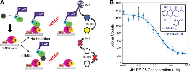
Intrinsic and acquired drug resistance and induction of secondary malignancies limit successful chemotherapy. Because mutagenic translesion synthesis (TLS) contributes to chemoresistance as well as treatment-induced mutations, targeting TLS is an attractive avenue for improving chemotherapeutics. However, development of small molecules with high specificity and in vivo efficacy for mutagenic TLS has been challenging. Here, we report the discovery of a small-molecule inhibitor, JH-RE-06, that disrupts mutagenic TLS by preventing recruitment of mutagenic POL ζ. Remarkably, JH-RE-06 targets a nearly featureless surface of REV1 that interacts with the REV7 subunit of POL ζ. Binding of JH-RE-06 induces REV1 dimerization, which blocks the REV1-REV7 interaction and POL ζ recruitment. JH-RE-06 inhibits mutagenic TLS and enhances cisplatin-induced toxicity in cultured human and mouse cell lines. Co-administration of JH-RE-06 with cisplatin suppresses the growth of xenograft human melanomas in mice, establishing a framework for developing TLS inhibitors as a novel class of chemotherapy adjuvants.

Keywords: POL ζ; REV1; REV7; chemoresistance; chemotherapy; cisplatin; translesion synthesis.


Conflict of interest statement

DECLARATION OF INTERESTS

P.Z. and J.H. are inventors of a patent on JH-RE-06. The remaining authors declare no competing interests.

Figures

Figure 1.
Figure 1.. Discovery and characterization of JH-RE-06, a small molecule inhibitor that blocks the REV1 CTD-REV7 interaction.
(A) A schematic overview of the ELISA assay. (B) Dose-dependent inhibition of the REV1 CTD-REV7 interaction by JH-RE-06 in the AlphaScreen™ assay. Fitting of the inhibition curve yields an IC50 value of 0.78 ± 0.16 μM for JH-RE-06. Error bars represent standard error of the mean (n=3).
Figure 2.
Figure 2.. Structural and biochemical characterization of the cREV1 CTD/JH-RE-06 complex.
(A) Structure of the cREV1 CTD/JH-RE-06 complex. Proteins are shown in the cartoon model, with protomer A colored in green and protomer B colored in cyan. JH-RE-06 is shown in the stick model, with the purple mesh representing the inhibitor omit map (2mFo-DFc) contoured at 1.0σ. (B) Surface representation of the cREV1 CTD/JH-RE-06 complex, illustrating the formation of a large ligand cavity at the dimeric REV1 CTD interface and the near encapsulation of JH-RE-06 within the cavity. (C) Interactions of JH-RE-06 with REV1 CTD residues. Denoted residue numbers correspond to the full-length REV1 protein. (D) Superimposition of the JH-RE-06-bound cREV1 CTD dimer (colored in green and cyan) with the POL κ RIR-REV1 CTD-REV7/3 translesionsome complex (colored in grey and pale cyan), illustrating the blockage of the REV1-REV7 interaction in the JH-RE-06-bound REV1 complex. (E) Binding of JH-RE-06 promotes the dimerization of the REV1 CTD in solution. Samples of the chimeric FLAG-tagged POL κ RIR-REV1 CTD in the presence of 0 μM or 100 μM JH-RE-06 were treated with increasing molar ratios of DSS and analyzed by SDS-PAGE followed by Western blotting with anti-FLAG antibody. Monomer and crosslinked dimer bands are labeled.
Figure 3.
Figure 3.. JH-RE-06 enhances cisplatin cytotoxicity and suppresses cisplatin-induced mutagenesis.
Cells were treated with DMSO or cisplatin (0.5 μM) for 24 hours, followed by JH-RE-06 (1.5 μM) treatment for additional 24 hours. Cells were washed and allowed to form colonies for 5–7 days and counted after staining with Coomassie brilliant blue R-250 stain. Shown in panels (A) HT1080 (human fibrosarcoma), (B) A375 (human melanoma), (C) KP (mouse KrasG12D;p53−/− lung adenocarcinoma), (D) LNCap (human prostate adenocarcinoma), and (E) AG01522 (human primary fibroblasts) are the relative colony forming ability of these cells in response to DMSO, JH-RE-06, cisplatin, and a combination dose of cisplatin and JH-RE-06. (F) The relative ability of HPRT+ HT1080 cells to mutate and form HPRT colonies in 6-TG media in the presence of DMSO, 1.5 μM JH-RE-06, 0.5 μM cisplatin, and the combination dose of 0.5 μM cisplatin and 1.5 μM JH-RE-06. Error bars represent standard error of the mean (n=6 for panels A-E and n=12 for panel F). Statistical analysis: one-way ANOVA with Tukey HSD post-hoc test. **P<0.01; N.S., not significant.
Figure 4.
Figure 4.. JH-RE-06 sensitizes cells to cisplatin and reduces HPRT mutations in a REV1-dependent manner.
The combination treatment of JH-RE-06 (1.5 μM) and cisplatin (0.5 μM) significantly reduced the colony forming ability in Rev1+/+ MEF cells (A), but not in Rev1−/− MEF cells (B), in comparison with cisplatin treatment alone. (C) Complementation of Rev1−/− MEF cells with a plasmid encoding REV1 by nucleofection fully restored the JH-RE-06 (1.5 μM) mediated sensitization to cisplatin (1 μM). siRNA knock-down of REV1 abolished JH-RE-06 (1.5 μM) mediated sensitization to cisplatin treatment (1 μM) in HT1080 (D) and A375 (E) cells. Treatment with JH-RE-06 (1.5 μM) significantly reduced spontaneous or cisplatin-induced (0.5 μM) HPRT mutation rates in Rev1+/+ MEF cells (F), but not in Rev1−/− MEF cells (G). Relative cell survival reflects the normalized colony forming ability of treated cells to DMSO controls. Error bars represent standard error of the mean (n=12 for panels A-C and F-G; n=6 for panels D–E). Statistical analysis: one-way ANOVA with Tukey HSD post-hoc test. **P<0.01; N.S., not significant.
Figure 5.
Figure 5.. JH-RE-06 improves tumor cell response to cisplatin in a xenograft mouse model.
(A) Inhibition of A375 xenograft tumor growth with (i) saline, (ii) JH-RE-06, (iii) cisplatin, and (iv) cisplatin and JH-RE-06. Compounds were formulated in 10% DMSO, 10% ethanol, 40% PEG-400, and 50% saline. The doses of JH-RE-06 and cisplatin per injection were 1.6 mg/kg and 1.0 mg/kg, respectively. Error bars represent standard error of the mean (n=10–12 xenograft tumors from 6 mice per treatment group). p-values for tumor volumes between each treatment group were calculated by the Welch’s t-test (Graphpad Prism). *p<0.05; N.S., not significant. (B) Survival curves of tumor-bearing mice treated with the four formulations above. Day 10 represents the first day of the specified drug administration (n=6 mice per treatment group in one representative experiment shown here, from a total of three independent experiments). A p-value of 0.0017 for the JH-RE-06 and cisplatin combination treatment vs. cisplatin treatment for survival studies was determined by using the Mantel-Cox log-rank test.

Comment in

References

    1. Actis ML, Ambaye ND, Evison BJ, Shao Y, Vanarotti M, Inoue A, McDonald ET, Kikuchi S, Heath R, Hara K, et al. (2016). Identification of the first small-molecule inhibitor of the REV7 DNA repair protein interaction. Bioorganic & medicinal chemistry 24, 4339–4346. - PMC - PubMed
    1. Adams PD, Grosse-Kunstleve RW, Hung LW, Ioerger TR, McCoy AJ, Moriarty NW, Read RJ, Sacchettini JC, Sauter NK, and Terwilliger TC (2002). PHENIX: building new software for automated crystallographic structure determination. Acta Crystallogr D Biol Crystallogr 58, 1948–1954. - PubMed
    1. Avkin S, Goldsmith M, Velasco-Miguel S, Geacintov N, Friedberg EC, and Livneh Z (2004). Quantitative analysis of translesion DNA synthesis across a benzo[a]pyrene-guanine adduct in mammalian cells: the role of DNA polymerase kappa. The Journal of biological chemistry 279, 53298–53305. - PubMed
    1. Baranovskiy AG, Lada AG, Siebler HM, Zhang Y, Pavlov YI, and Tahirov TH (2012). DNA polymerase delta and zeta switch by sharing accessory subunits of DNA polymerase delta. The Journal of biological chemistry 287, 17281–17287. - PMC - PubMed
    1. Bhat A, Wu Z, Maher VM, McCormick JJ, and Xiao W (2015). Rev7/Mad2B plays a critical role in the assembly of a functional mitotic spindle. Cell Cycle 14, 3929–3938. - PMC - PubMed

Publication types

MeSH terms