. 2019 May 24:13:403.
doi: 10.3389/fnins.2019.00403. eCollection 2019.

L-Lactate Promotes Adult Hippocampal Neurogenesis

Affiliations

L-Lactate Promotes Adult Hippocampal Neurogenesis

Yaeli Lev-Vachnish et al. Front Neurosci. .

Abstract

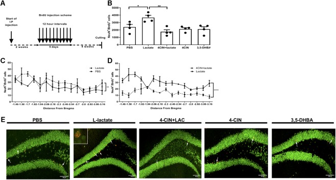
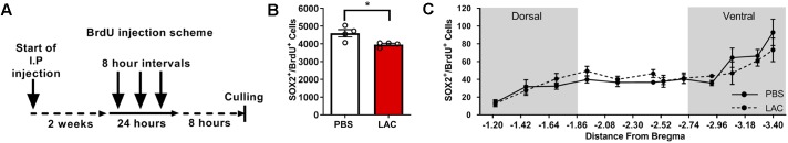
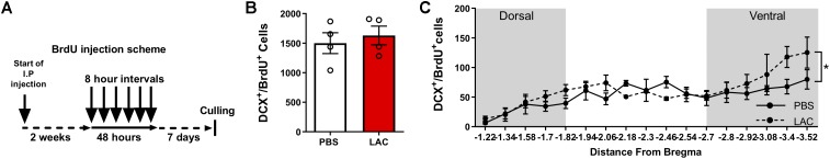
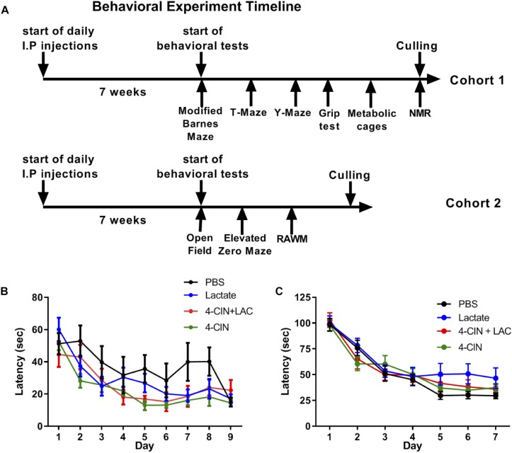
Neurogenesis, the formation of new neurons in the adult brain, is important for memory formation and extinction. One of the most studied external interventions that affect the rate of adult neurogenesis is physical exercise. Physical exercise promotes adult neurogenesis via several factors including brain-derived neurotrophic factor (BDNF) and vascular endothelial growth factor (VEGF). Here, we identified L-lactate, a physical exercise-induced metabolite, as a factor that promotes adult hippocampal neurogenesis. While prolonged exposure to L-lactate promoted neurogenesis, no beneficial effect was exerted on cognitive learning and memory. Systemic pharmacological blocking of monocarboxylate transporter 2 (MCT2), which transports L-lactate to the brain, prevented lactate-induced neurogenesis, while 3,5-dihydroxybenzoic acid (3,5-DHBA), an agonist for the lactate-receptor hydroxycarboxylic acid receptor 1 (HCAR1), did not affect adult neurogenesis. These data suggest that L-lactate partially mediates the effect of physical exercise on adult neurogenesis, but not cognition, in a MCT2-dependent manner.

Keywords: L-lactate; MCT2; hippocampus; neurogenesis; neuronal progenitor cells.


Figures

Figure 1
Figure 1
Exogenous L-lactate increases the number of NeuN+BrdU+ cells in the dentate gyrus. (A) Experiment design: Mice were i.p. injected with L-lactate, PBS, 4-CIN and 4-CIN, followed by L-lactate or 3,5-DHBA (n = 4 per treatment group), for 2 weeks prior to receiving 10 consecutive BrdU injections at 12 h intervals and sacrificed 4 weeks after the last injection. (B) Number of NeuN+/BrdU+ in the DG: NeuN+/BrdU+ cells were counted in the DG and the total DG population estimation was extrapolated for each animal. Results were averaged across treatment group. Number of NeuN+/BrdU+ cells in L-lactate-treated mice was significantly higher compared to PBS (P < 0.05) and 4-CIN + L-lactate (∗∗P < 0.01). (C) NeuN+/BrdU+ averaged cell distribution according to the distance from Bregma position. NeuN+/BrdU+ cell distribution was significantly higher in the L-lactate group compared with PBS treatment (∗∗p < 0.01) or (D) 4-CIN + L-lactate (∗∗∗∗p < 0.0001). (E) Representative NeuN+/BrdU+ cells in the SGZ of PBS, L-lactate, 4-CIN + L-lactate, 4-CIN, and 3,5-DHBA injected mice.
Figure 2
Figure 2
Exogenous L-lactate reduced the number of Sox2+BrdU+ cells in the dentate. (A) Experiment design: Mice were injected for 2 weeks with L-lactate (1.75 g/kg, n = 4) or PBS (n = 4) and were then injected with BrdU (100 mg/kg) three times at 8 h intervals and sacrificed 8 h after the last injection. (B) Number of Sox2+/BrdU+ in the DG; Sox2+/BrdU+ cells were counted for each experimental animal’s DG and total DG population estimation was extrapolated. Results were averaged across treatment group. L-lactate treatment reduces the number of Sox2+/BrdU+ cells (p < 0.05). (C) Sox2+/BrdU+ average cell distribution according to the distance from Bregma position. No significant differences were found between L-lactate and PBS throughout the DG (P > 0.05).
Figure 3
Figure 3
Exogenous L-lactate does not affect the number of DCX+BrdU+ cells in the dentate gyrus. (A) Experiment design: Mice were i.p. injected with 1.75 g/kg L-lactate solution (n = 4) or PBS (n = 4) and were then injected with BrdU for 2 days at 8 h intervals and sacrificed 5 days after the last injection. (B) Total number of DCX+/BrdU+ cells in the DG; DCX+/BrdU+ cells were counted for each experimental animal’s DG and total DG population estimation was extrapolated. Results were averaged across treatment group. No significant differences were found between L-lactate and PBS throughout the DG (P > 0.05). (C) DCX+/BrdU+ averaged cell distribution according to the distance from Bregma position. DCX+/BrdU+ cell distribution was significantly higher in L-lactate group compared with PBS treatment (p < 0.01).
Figure 4
Figure 4
Chronic L-lactate administration does not affect long-term hippocampal-dependent spatial learning. (A) Experiment design: Mice were i.p. injected with L-lactate, PBS, 4-CIN and 4-CIN followed by L-lactate (n = 8–12 per group) for 7 weeks before starting behavioral experiments for nine additional weeks. During the behavioral experiments, treatments were continuously administered. Cohort 1 (upper timeline) was tested in the Barnes maze, T-maze, Y-maze, grip strength test, metabolic cages and finally underwent NMR analysis for body composition. Cohort 2 was tested in the open field arena, the elevated zero maze and the radial arm water maze. (B) Mice were tested using the RAWM; latency to reach platform. No significant effect was found between treatments (P > 0.05). (C) Mice were tested using the modified Barnes maze; latency to reach target hole. No significant effect was found between treatments (P > 0.05).

References

    1. Afzalpour M. E., Chadorneshin H. T., Foadoddini M., Eivari H. A. (2015). Comparing interval and continuous exercise training regimens on neurotrophic factors in rat brain. Physiol. Behav. 147 78–83. 10.1016/j.physbeh.2015.04.012 - DOI - PubMed
    1. Alvarez Z., Castano O., Castells A. A., Mateos-Timoneda M. A., Planell J. A., Engel E., et al. (2014). Neurogenesis and vascularization of the damaged brain using a lactate-releasing biomimetic scaffold. Biomaterials 35 4769–4781. 10.1016/j.biomaterials.2014.02.051 - DOI - PubMed
    1. Baptista P., Andrade J. P. (2018). Adult hippocampal neurogenesis: regulation and possible functional and clinical correlates. Front. Neuroanat. 12:44. 10.3389/fnana.2018.00044 - DOI - PMC - PubMed
    1. Barak B., Feldman N., Okun E. (2015). Cardiovascular fitness and cognitive spatial learning in rodents and in humans. J. Gerontol. A Biol. Sci. Med. Sci. 70 1059–1066. 10.1093/gerona/glu162 - DOI - PMC - PubMed
    1. Barros L. F. (2013). Metabolic signaling by lactate in the brain. Trends Neurosci. 36 396–404. 10.1016/j.tins.2013.04.002 - DOI - PubMed

LinkOut - more resources