Review
. 2019 May 24:10:1084.
doi: 10.3389/fimmu.2019.01084. eCollection 2019.

Macrophage Polarization: Different Gene Signatures in M1(LPS+) vs. Classically and M2(LPS-) vs. Alternatively Activated Macrophages

Affiliations
Review

Macrophage Polarization: Different Gene Signatures in M1(LPS+) vs. Classically and M2(LPS-) vs. Alternatively Activated Macrophages

Marco Orecchioni et al. Front Immunol. .

Erratum in

Abstract

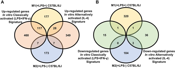
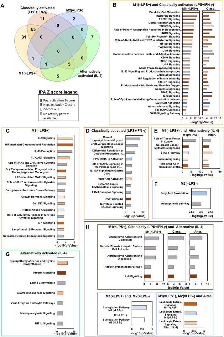
Macrophages are found in tissues, body cavities, and mucosal surfaces. Most tissue macrophages are seeded in the early embryo before definitive hematopoiesis is established. Others are derived from blood monocytes. The macrophage lineage diversification and plasticity are key aspects of their functionality. Macrophages can also be generated from monocytes in vitro and undergo classical (LPS+IFN-γ) or alternative (IL-4) activation. In vivo, macrophages with different polarization and different activation markers coexist in tissues. Certain mouse strains preferentially promote T-helper-1 (Th1) responses and others Th2 responses. Their macrophages preferentially induce iNOS or arginase and have been called M1 and M2, respectively. In many publications, M1 and classically activated and M2 and alternatively activated are used interchangeably. We tested whether this is justified by comparing the gene lists positively [M1(=LPS+)] or negatively [M2(=LPS-)] correlated with the ratio of IL-12 and arginase 1 in transcriptomes of LPS-treated peritoneal macrophages with in vitro classically (LPS, IFN-γ) vs. alternatively activated (IL-4) bone marrow derived macrophages, both from published datasets. Although there is some overlap between in vivo M1(=LPS+) and in vitro classically activated (LPS+IFN-γ) and in vivo M2(=LPS-) and in vitro alternatively activated macrophages, many more genes are regulated in opposite or unrelated ways. Thus, M1(=LPS+) macrophages are not equivalent to classically activated, and M2(=LPS-) macrophages are not equivalent to alternatively activated macrophages. This fundamental discrepancy explains why most surface markers identified on in vitro generated macrophages do not translate to the in vivo situation. Valid in vivo M1/M2 surface markers remain to be discovered.

Keywords: M1; M2; cancer; innate immunity; macrophage.


Figures

Figure 1
Figure 1
Signature comparison for C57BL/6 macrophages. (A) Venn diagram showing overlap between genes whose expression was positively [M1(=LPS+)C57BL/6J] or negatively [M2(=LPS–)C57BL/6J] correlated with IL12/arginase ratio in vivo with genes upregulated in vitro classically activated (LPS+IFN-γ) macrophages (left) or in vitro alternatively activated (IL-4) (right) macrophages vs. unstimulated. (B) Venn diagram showing overlap between genes whose expression was positively [M1(=LPS+)C57BL/6J] or negatively [M2(=LPS–)C57BL/6J] correlated with IL12/arginase in vivo with genes downregulated in vitro classically activated (LPS+IFN-γ) macrophages (left) or in vitro alternatively activated (IL-4) (right) macrophages vs. unstimulated.
Figure 2
Figure 2
Ingenuity pathways analysis in vivo M1(=LPS+)C57BL/6J, M2(=LPS–)C57BL/6J, and in vitro classically (LPS+IFN-γ) or alternatively activated (IL-4) macrophages. (A) Venn diagram showing the key canonical pathways enriched in vivo M1(=LPS+)C57BL/6J, M2(=LPS–)C57BL/6J, M2(=LPS–)C57BL/6J, classically activated (LPS+IFN-γ) and alternatively activated (IL-4) macrophage signatures as determined by Ingenuity pathway analysis (IPA). The number of pathways is sorted by a P-value cutoff of 0.001. (B–H) Selected canonical pathways ranked based on –log(P-value) divided as reported in the Venn diagram are shown in the boxes. The Z-score of each pathway is reported by the color of the bars (see legend). Light/Dark shades represent smaller/larger absolute values of Z-score.
Figure 3
Figure 3
In vivo macrophage signatures predict survival in osteosarcoma cancer biopsy transcriptomes. Survival data for human osteosarcoma cancer biopsies (GSE21257) were analyzed for the impact of M1(=LPS+) and classically activated (LPS+IFN-γ) (A) and M2(=LPS−) and alternatively activated (IL-4) (B) gene expression signatures in the tumor biopsy transcriptome. Kaplan–Meier curves were plotted using ProggeneV2, divided by the median of the mean expression of a tumor-specific gene list (in boxes). Hazard ratio (HR, cox proportional hazard analysis) and significance (log rank P-value) are shown. Red, green curves indicate high, low expression of the respective signature genes. The two vertical black lines indicates 3 and 5 years, respectively.

References

    1. Ginhoux F, Guilliams M. Tissue-resident macrophage ontogeny and homeostasis. Immunity. (2016) 44 439–49. 10.1016/j.immuni.2016.02.024 - DOI - PubMed
    1. van Furth R, Cohn ZA. The origin and kinetics of mononuclear phagocytes. J Exp Med. (1968) 128:415–35. 10.1084/jem.128.3.415 - DOI - PMC - PubMed
    1. Fogg DK, Sibon C, Miled C, Jung S, Aucouturier P, Littman DR, et al. . A clonogenic bone marrow progenitor specific for macrophages and dendritic cells. Science. (2006) 311:83–7. 10.1126/science.1117729 - DOI - PubMed
    1. Sathe P, Metcalf D, Vremec D, Naik SH, Langdon WY, Huntington ND, et al. Lymphoid tissue and plasmacytoid dendritic cells and macrophages do not share a common macrophage-dendritic cell-restricted progenitor. Immunity. (2014) 41:104–15. 10.1016/j.immuni.2014.05.020 - DOI - PubMed
    1. Lavin Y, Mortha A, Rahman A, Merad M. Regulation of macrophage development and function in peripheral tissues. Nat Rev Immunol. (2015) 15:731–44. 10.1038/nri3920 - DOI - PMC - PubMed

Publication types