. 2019 Apr 3;6(11):1900034.
doi: 10.1002/advs.201900034. eCollection 2019 Jun 5.

A Genomic, Evolutionary, and Mechanistic Study of MCR-5 Action Suggests Functional Unification across the MCR Family of Colistin Resistance

Affiliations

A Genomic, Evolutionary, and Mechanistic Study of MCR-5 Action Suggests Functional Unification across the MCR Family of Colistin Resistance

Huimin Zhang et al. Adv Sci (Weinh). .

Abstract

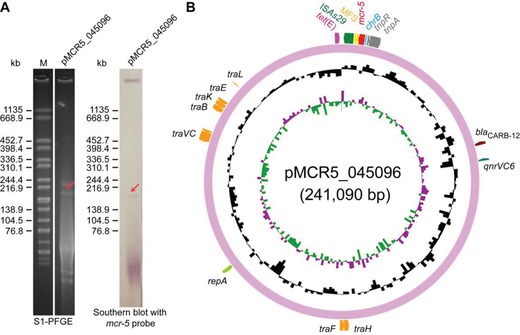
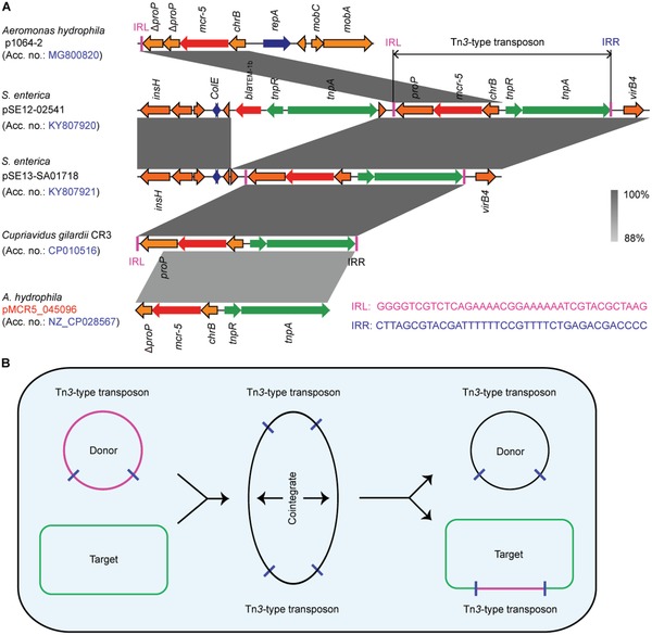
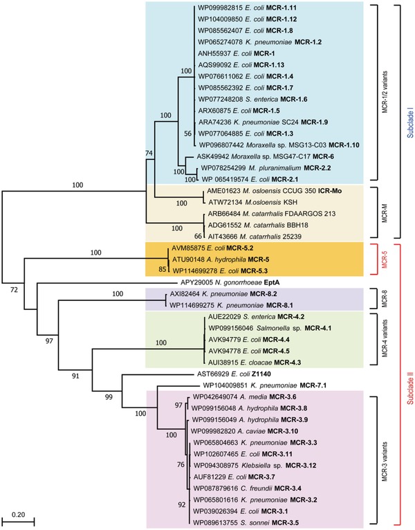
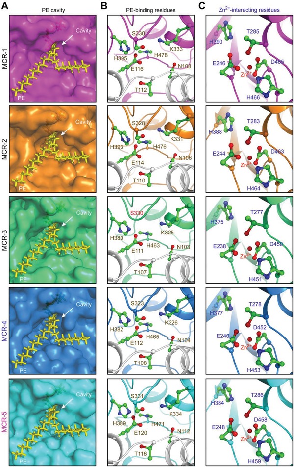
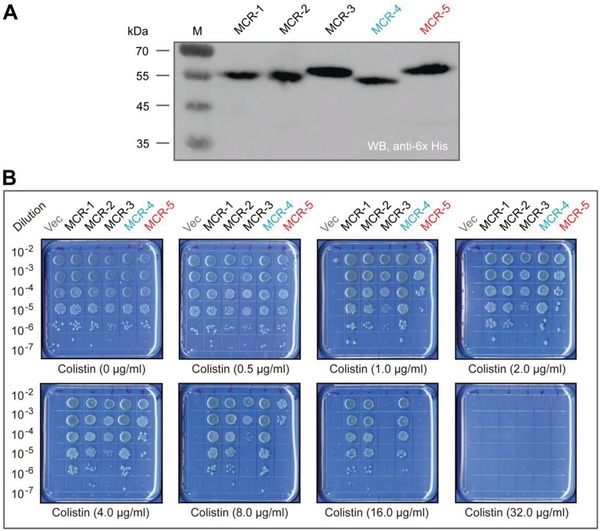
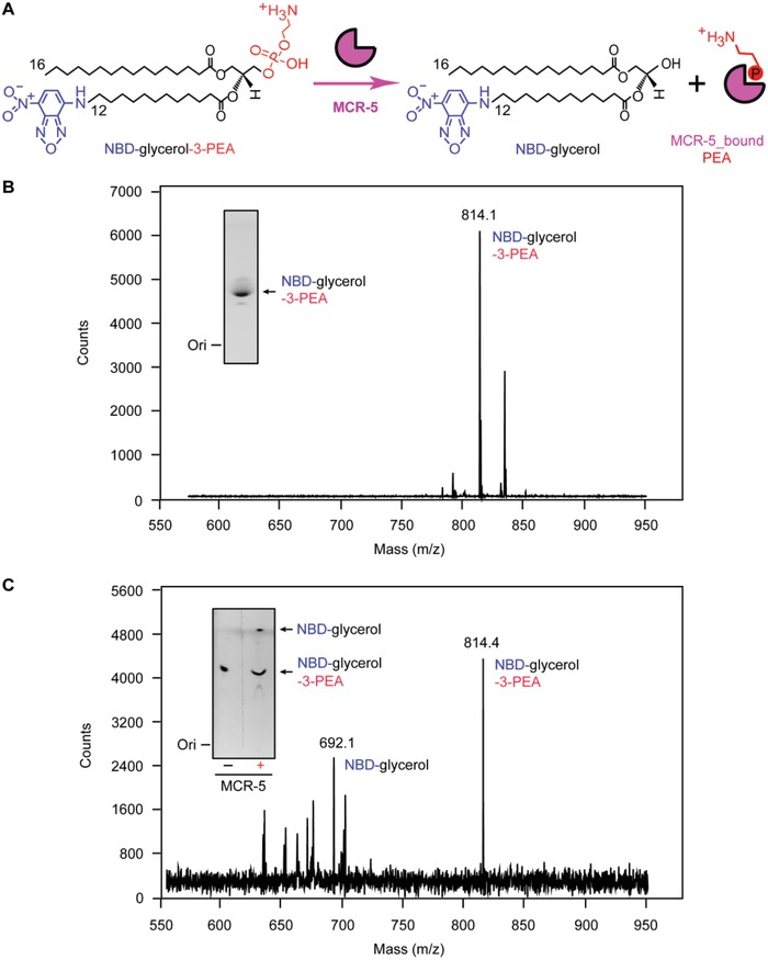
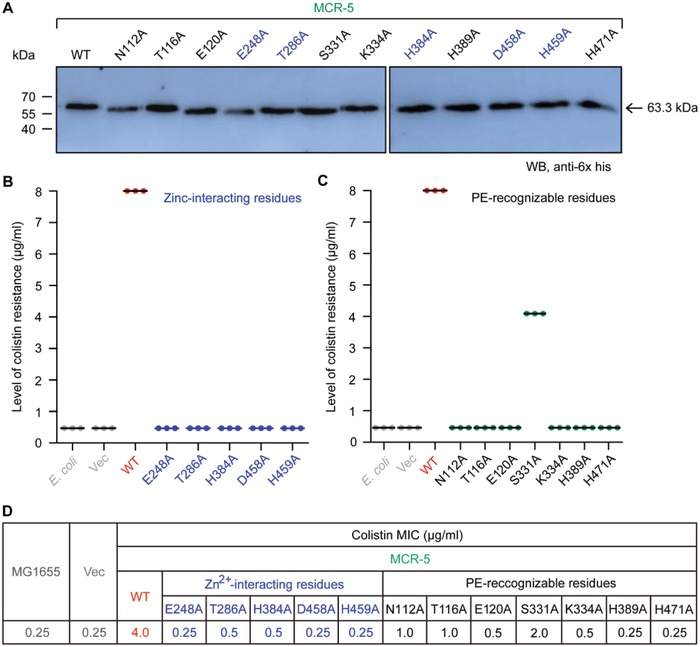
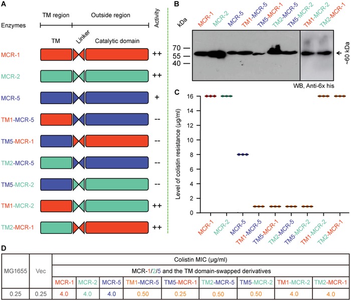
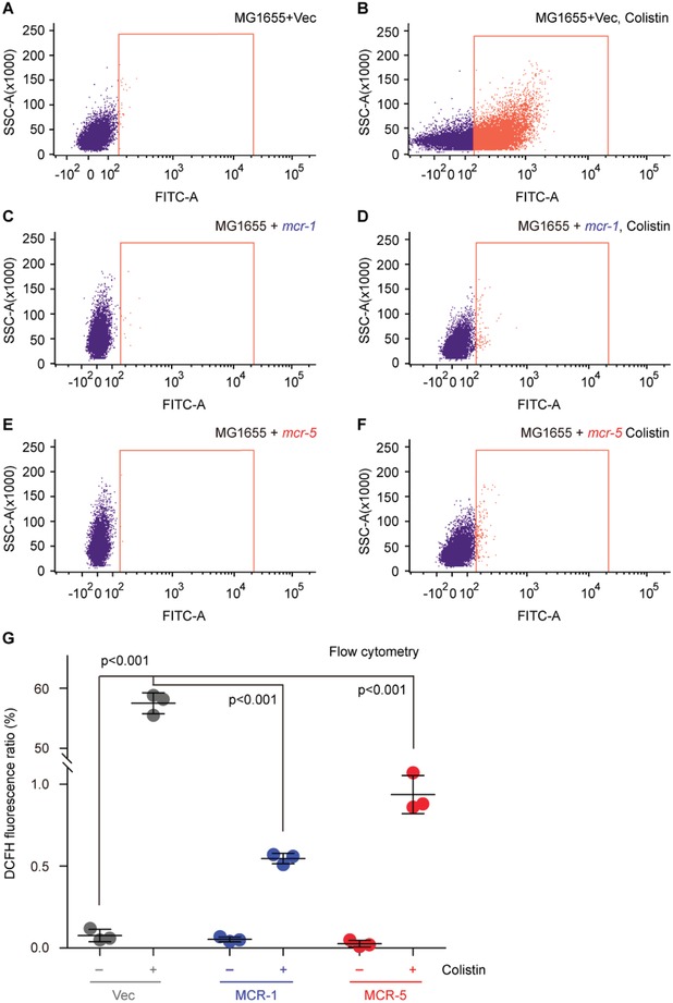
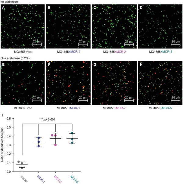
A growing number of mobile colistin resistance (MCR) proteins is threatening the renewed interest of colistin as a "last-resort" defense against carbapenem-resistant pathogens. Here, the comparative genomics of a large plasmid harboring mcr-5 from Aeromonas hydrophila and the structural/functional perspectives of MCR-5 action are reported. Whole genome sequencing has identified the loss of certain parts of the Tn3-type transposon typically associated with mcr-5, providing a clue toward its mobilization. Phylogeny of MCR-5 suggests that it is distinct from the MCR-1/2 sub-lineage, but might share a common ancestor of MCR-3/4. Domain-swapping analysis of MCR-5 elucidates that its two structural motifs (transmembrane domain and catalytic domain) are incompatible with its counterparts in MCR-1/2. Like the rest of the MCR family, MCR-5 exhibits a series of conservative features, including zinc-dependent active sites, phosphatidylethanolamine-binding cavity, and the mechanism of enzymatic action. In vitro and in vivo evidence that MCR-5 catalyzes the addition of phosphoethanolamine to the suggestive 4'-phosphate of lipid A moieties is integrated, and results in the consequent polymyxin resistance. In addition, MCR-5 alleviates the colistin-induced formation of reactive oxygen species in E. coli. Taken together, the finding suggests that a growing body of MCR family resistance enzymes are functionally unified.

Keywords: Aeromonas hydrophila; MCR‐5; colistin resistance; functional unification; lipid A; phosphatidylethanolamine (PE) cavity; ping‐pong reaction mechanism; transferable resistance.


Conflict of interest statement

The authors declare no conflict of interest.

Figures

Figure 1
Figure 1
A new mcr‐5‐harboring plasmid from Aeromonas hydrophila. A) Use of Southern blot to estimate the size of the newly identified mcr‐5‐harboring plasmid pMCR‐5_045096 following the separation with SI‐PFGE. B) Circular illustration for genomic map of pMCR‐5_045096. mcr‐5 is indicated in red.
Figure 2
Figure 2
Genomic context of mcr‐5‐containing plasmids and genetic analyses for mcr‐5 dissemination. A) Colinear analyses for genetic environment of mcr‐5‐neighboring loci from different plasmid reservoirs or chromosome. Linear comparison of the mcr5‐carrying plasmids p1064‐2 (MG800820), pSE12‐02541 (KY807920), pSE13‐SA01718 (KY807921), chromosome fragment of C. gilardii strain CR3, and plasmid pMCR5_045096 (CP028567) was performed in this study. Boxed arrows represent the position and transcriptional direction of ORFs. Regions of >99% identity are marked by gray shading. Genes associated with replication associated genes are colored dark blue, antibiotic resistance genes are colored red, insertion sequences are colored green, and other genes are colored orange. IRL, terminal inverted repeats of left. IRR, terminal inverted repeats of right. B) Scheme for the replicative transposition cycle of Tn3‐type transposons harboring mcr‐5. The black circle represents the donor ColE‐like plasmids carrying mcr‐5‐Tn3‐type transposons. The rectangle represents the target of Tn3‐type transposons.
Figure 3
Figure 3
Phylogeny of MCR‐5. An unrooted phylogenetic tree of MCR‐5 and its close homologs is presented with two distinct subclades (Subclade I and Subclade II) with paraphyletic branches. Full protein sequences of MCR enzymes were applied to generate phylogenic tree. Subclade I contains MCR‐1/2 variants (light pink) and its progenitors MCR‐M (light orange), whereas Subclade II comprises MCR‐5 (in yellow), MCR‐4 variants (light blue), and MCR‐3 variants (in green). MCR‐5 is highlighted in bold red font. Abbreviations: E. coli, Escherichia coli; K. pneumoniae, Klebsiella pneumoniae; S. enterica, Salmonella enterica; M. pluranimalium, Moraxella pluranimalium; M. osloensis, Moraxella osloensis; M. catarrhalis, Moraxella catarrhalis; N. gonorrhoeae, Neisseria gonorrhoeae; A. media, Aeromonas media; A. hydrophila, Aeromonas hydrophila; A. caviae, Aeromonas caviae; C. freundii, Citrobacer freundii; S. sonnei, Shigella sonnei.
Figure 4
Figure 4
Parallels among PE‐recognizable cavities of the MCR family of lipid A modifiers. A) Similarity of PE cavities in different members of MCR family. B) Visualization for a conserved motif comprising 7 PE‐binding residues. C) Parallels in zinc‐binding residues of MCR‐5 and MCR‐1/2/3/4. PyMol is applied to generate the photographs of surface structure, PE cavities, and enzymatically catalytic center.
Figure 5
Figure 5
Comparative analyses for colistin resistance levels in E. coli conferred by an array of different MCR versions. A) Western blot‐aided comparative analyses of functional expression of mcr‐1/2/3/4/5 in vivo. B) Growth viability of E. coli harboring different version of MCR family of enzymes on the LBA plates supplied with varied level of colistin. A representative result is given from three independent experiments. Designation: Vec, pBAD24; WB, Western blot.
Figure 6
Figure 6
Enzymatic action of MCR‐5 in vitro. A) Scheme for chemical reaction of MCR‐5 in hydrolyzing an alternative lipid substrate of PE, NBD‐glycerol‐3‐PEA, into NBD‐glycerol and an adduct of MCR‐5_bound PEA. B) LC/MS identity of the alternative lipid substrate of PE, NBD‐glycerol‐3‐PEA. C) LC/MS‐based detection for the mixture of MCR‐5 reaction with NBD‐glycerol‐3‐PEA as substrate. Inside gel separately refers to TLC assays for the substrate of NBD‐glycerol‐3‐PEA (B) and its resultant product NBD‐glycerol (C).
Figure 7
Figure 7
Mapping genetic elements necessary for MCR‐5 colistin resistance. A) Western blot‐based expression assays for MCR‐5 and its 12‐point mutants in E. coli. B) Site‐directed mutagenesis analyses for the Zn2+‐binding motif of MCR‐5 in the context of colistin resistance using the colistin susceptibility tests. The five residues in Zn2+‐binding motif of MCR‐5 denote E248, T286, H384, D458, and H459, respectively. C) Colistin susceptibility‐based dissection of the PE‐interactive residues of MCR‐5. The seven residues denote N112, T116, E120, S331, K334, H389, and H471, respectively. Assays of three individual bacterial viability on colistin agar plates were conducted. D) Minimum inhibitory concentration (MIC) of colistin of E. coli harboring mcr‐5 and/or its point mutants. Designation: Vec, pBAD24; WT, wild‐type.
Figure 8
Figure 8
Domain‐swapping analyses of MCR‐1, MCR‐2, and MCR‐5. A) Scheme for domain‐swapped constructs between MCR‐5 and MCR‐1/2. B) Western blot‐based confirmation for functional expression of mcr‐5 and its hybrid versions in E. coli. C) Bacterial viability of E. coli expressing mcr‐5 and its hybrid derivatives on the LBA plates supplied with colistin. Three independent tests were performed. D) Colistin MIC of E. coli MG1655 harboring the wild‐type of mcr‐5 or its hybrid derivatives. In total, six derivatives from domain‐swapping among MCR‐1, MCR‐2, and MCR‐5. Designations: Vec, pBAD24; TM1‐MCR‐5, a derivative of MCR‐5 with TM1 region of MCR‐1 in place of its native TM domain; TM5‐MCR‐1, a hybrid version of MCR‐1 whose TM region is replaced with the counterpart in MCR‐5; TM2‐MCR‐5, a mosaic version of MCR‐5 whose TM region is exchanged with that of MCR‐2; TM5‐MCR‐2, a hybrid derivative of MCR‐2 whose TM region is replaced with that of MCR‐5; TM1‐MCR‐2, a hybrid derivative of MCR‐2 whose TM region is replaced with that of MCR‐1; and TM2‐MCR‐1, a derivative of MCR‐1 whose TM region is replaced with that of MCR‐2.
Figure 9
Figure 9
FACS analyses of colistin‐induced ROS level in E. coli. A,B) The colistin treatment boosts the accumulation of ROS in E. coli with empty vector. C,D) The presence of colistin cannot promote efficient formation of ROS in mcr‐1‐harboring in E. coli. E,F) The expression of MCR‐5 catalyzes the attachment of PEA to the suggestive 4′‐phosphate position of lipid A anchored on E. coli surface and prevents efficient production of intracellular ROS. G) Use of flow cytometry to measure the relative level of ROS in E. coli alone or carrying mcr‐1/5. Flow cytometry of ROS was performed with a BD FACSVerse flow cytometer in which around 10 000 cells are counted at a flow rate of 35 mL min−1. The fluorescence of the dye DCFH2‐DA was excited with a 488 nm argon laser and emission was detected with the FL1 emission filter at 525 nm using FL1 photomultiplier tub. The minus symbol denotes the absence of colistin, and the plus symbol refers to the addition of colistin. The data were expressed using one‐way analysis of variance (ANOVA) followed by Tukey–Kramer multiple comparisons post hoc test.47 Statistical significance was set at p < 0.001. Designations: Vec, pBAD24; “−,” no addition of colistin; “+,” addition of colistin.
Figure 10
Figure 10
Functional expression of mcr‐1/2/5 genes is accompanied with bacterial metabolic fitness. Regardless of the presence of the A,E) empty vector pBAD24 or B–D) MCR‐1/2/5, no addition of the inducer arabinose cannot significantly alter bacterial survival in E. coli MG1655. F–H) Confocal microscopy assays illustrate that arabinose (0.2%)‐triggered expression of MCR‐1/2/5 interferes bacterial viability. I) Measurement of the relative ratio of LIVE/DEAD E. coli strains expressing MCR‐1/2/5. 0.2% (w/v) l‐arabinose was added to initiate the expression of mcr‐1/2/5. Bacterial cells were stained with LIVE/DEAD kit, giving the images with confocal laser scanning microscopy. Green and red refer to live and dead cell. Vector refers to pET21. The data were expressed using one‐way analysis of variance (ANOVA) followed by Tukey–Kramer multiple comparisons post hoc test.47 Statistical significance was set at ***p < 0.001.

References

    1. Ferri M., Ranucci E., Romagnoli P., Giaccone V., Crit. Rev. Food Sci. Nutr. 2017, 57, 2857. - PubMed
    1. de Kraker M. E., Stewardson A. J., Harbarth S., PLoS Med. 2016, 13, e1002184. - PMC - PubMed
    1. WHO , 2014.
    1. Li J., Nation R. L., Turnidge J. D., Milne R. W., Coulthard K., Rayner C. R., Paterson D. L., Lancet Infect. Dis. 2006, 6, 589. - PubMed
    1. Poirel N. K. L., Liassine N., Thanh D., Nordmann P., Lancet Infect. Dis. 2016, 16, 281. - PubMed

LinkOut - more resources