. 2019 Apr 23:2019:4328219.
doi: 10.1155/2019/4328219. eCollection 2019.

Novel Hybrid Gels Made of High and Low Molecular Weight Hyaluronic Acid Induce Proliferation and Reduce Inflammation in an Osteoarthritis In Vitro Model Based on Human Synoviocytes and Chondrocytes

Affiliations

Novel Hybrid Gels Made of High and Low Molecular Weight Hyaluronic Acid Induce Proliferation and Reduce Inflammation in an Osteoarthritis In Vitro Model Based on Human Synoviocytes and Chondrocytes

Antonietta Stellavato et al. Biomed Res Int. .

Erratum in

Retraction in

Abstract

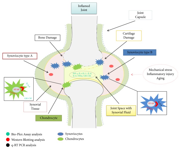
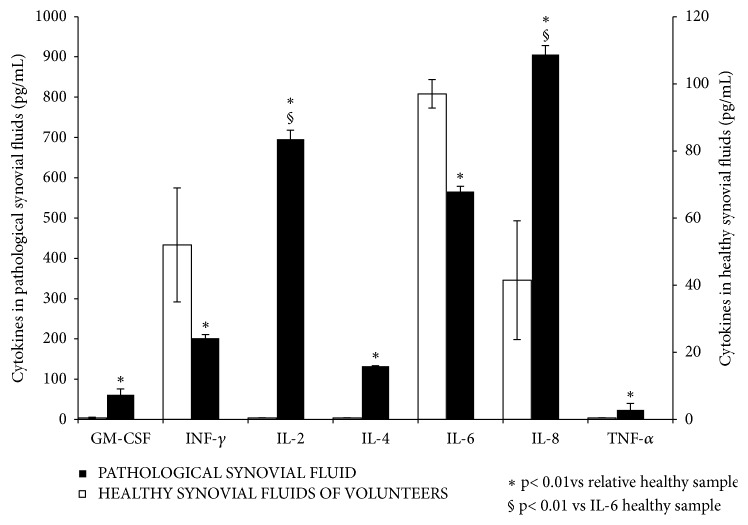
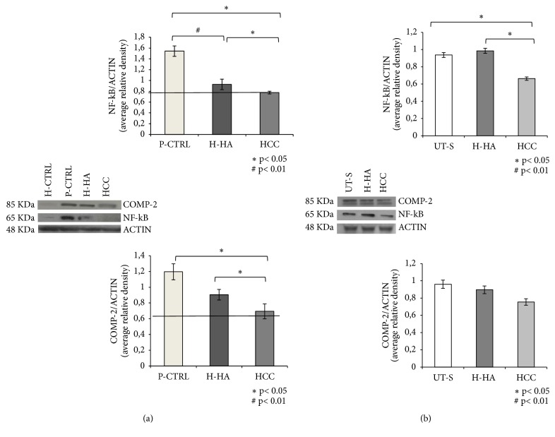
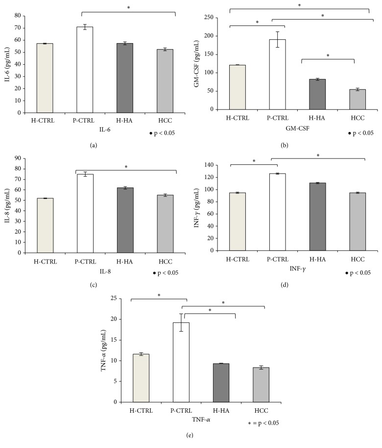
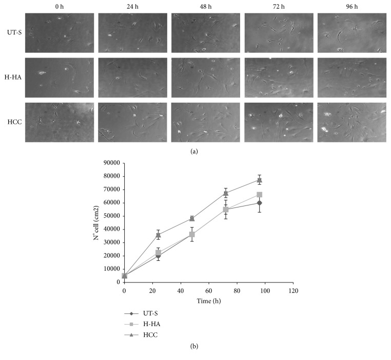
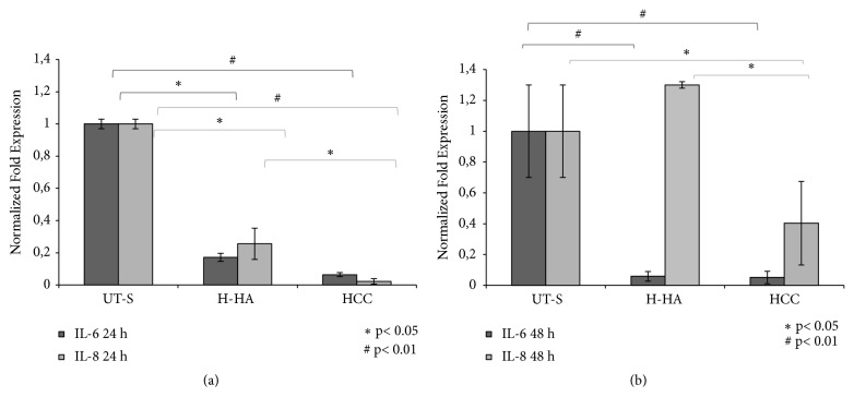
High molecular weight hyaluronan (H-HA) has a pivotal role in the maintenance of normal functions of synovial fluid and structure of the articular joint, but it has been shown that its concentration is reduced in patients affected by degenerative cartilage diseases, such as osteoarthritis (OA). The aim of this study was to investigate the anti-inflammatory effects and properties of hybrid cooperative complexes based on high and low molecular weight hyaluronan (HCC) compared to H-HA on human primary cells derived by pathological joints. In addition, the rheological behavior of HCC was evaluated in order to define their potential as viscosupplement gel in degenerated joints. The experiments were performed using an in vitro model of OA based on human chondrocytes and synoviocytes isolated from degenerated joints of patients hospitalized for surgical replacement. In order to assess the anti-inflammatory effects of HCC, we evaluated NF-kB, COMP-2, IL-6, and IL-8 as specific markers at the transcriptional and/or protein level. Moreover, the proliferative properties of HCC were assessed using time lapse video microscopy. We showed that chondrocytes and synoviocytes clearly presented an altered cytokine profile compatible with a severe ongoing inflammation status. H-HA and, above all, HCC significantly reduced levels of the specific biomarkers evaluated and improved cartilage healing. The rheological profile indicated HCC suitability for intra-articular injection in joint diseases. HCC viscoelastic properties and the protective/anti-inflammatory effect on human chondrocytes and synoviocytes suggest the novel HCC-based gels as a valid support for OA management.


Figures

Figure 1
Figure 1
Schematic resume of signaling investigated with related techniques used; in particular, synovial fluid and type B chondrocytes and synoviocytes have been objects of our studies. Gene expression and protein production were analyzed to assess the inflammation status in OA joints and then in derived cell populations.
Figure 2
Figure 2
HCC rheological characterization. (a) HCC dynamic viscosity as a function of the shear rate. (b) Dynamic Moduli for HCC as a function of the frequency.
Figure 3
Figure 3
Analysis of synovial fluids of four patients affected by OA by the Pro-Human Cytokine Multiplex Assay. The levels of the following cytokines were analyzed; GM-CSF, INF-γ, IL-2, IL-4, IL-6, IL-8, and TNF-α (pg/mL). The results are reported as the average of values for each cytokine assessed in every sample compared to the average of the cytokine levels in healthy fluids. Data presented by Tsuchida et al. (2014) [17] and Papalia et al. (2016) [26] and here presented to easily obtain a direct comparison. T-test analyses were performed between each pathological cytokine with respect to their healthy control (p<0.01) in addition to pathological IL-2 and IL-8 levels with respect to healthy IL-6 level (§p<0.01).
Figure 4
Figure 4
WB analysis of NF-kB and COMP-2 protein levels in chondrocytes (a) and synoviocytes. (b) Densitometric analysis of specifics bands obtained for NF-kB and COMP-2 expression has been normalized with respect to actin expression. Data shown are an average of two different western blotting with two densitometric recording each, reported as mean values ± SD. (a) Significant differences between H-HA and HCC versus P-CTRL and H-HA versus HCC are indicated with asterisks p<0.05, #p<0.01. Data shown are means ± SD. (b) Significant differences between H-HA and HCC versus P-CTRL and H-HA versus HCC are indicated with asterisks p<0.05, #p<0.01.
Figure 5
Figure 5
Analysis of IL-6, GM-CSF, IL-8, INF-γ, and TNF-α levels in supernatants (pg/mL) by the Bio-Plex cytokines assay of chondrocytes treated for 48 h with H-HA or HCC 0.32% w/v with respect to H-CTRL and P-CTRL. Data shown are means ± SD. Significant differences between H-HA and HCC versus P-CTRL, H-HA versus HCC, and H-HA or HCC versus H-CTRL are indicated with asterisks p<0.05, #p<0.01.
Figure 6
Figure 6
Human synoviocytes proliferation assay. (a) Pictures of cells at experimental times: 24, 48, 72, and 96 h; the proliferative properties of HCC were compared to the single H-HA and untreated synoviocytes (UT-S). (b) Cell growth curves were obtained through Image-Pro Plus 1.5 software (Media Cybernetics).
Figure 7
Figure 7
mRNA expression analysis of IL-6 and IL-8 in human synoviocytes isolated from osteoarthritic synovial fluid through qRT-PCR. (a) Quantitative gene expression of IL-6 and IL-8 in primary synoviocytes after 24 h of treatment with H-HA and HCC 0.32% w/v with respect to UT-S. (b) Gene expression of IL-6 and IL-8 in synoviocytes after 48 h of treatment with H-HA and HCC 0.32% w/v with respect to UT-S. Data shown are means ± SD. Significant differences between H-HA and HCC versus UT-S and H-HA versus HCC are indicated with asterisks p<0.05, #p<0.01.

References

    1. Glyn-Jones S., Palmer A. J., Agricola R., et al. Osteoarthritis. The Lancet. 2015;386(9991):376–387. doi: 10.1016/S0140-6736(14)60802-3. - DOI - PubMed
    1. Calamia V., Fernàndez-Puente P., Mateos J., et al. Pharmacoproteomic study of three different chondroitin sulfate compounds on intracellular and extracellular human choncrocyte proteomes. Molecular & Cellular Proteomics. 2012;11(6):p. M111.013417 - PMC - PubMed
    1. Stellavato A., Tirino V., de Novellis F., et al. Biotechnological chondroitin a novel glycosamminoglycan with remarkable biological function on human primary chondrocytes. Journal of Cellular Biochemistry. 2016;9999:1–12. doi: 10.1002/jcb.25556. - DOI - PMC - PubMed
    1. Hiligsmann M., Cooper C., Arden N., et al. Health economics in the field of osteoarthritis: an expert's consensus paper from the european society for clinical and economic aspects of osteoporosis and osteoarthritis (ESCEO) Seminars in Arthritis and Rheumatism. 2013;43(3):303–313. doi: 10.1016/j.semarthrit.2013.07.003. - DOI - PubMed
    1. Huang D., Zhao Q., Liu H., Guo Y., Xu H. PPAR-α agonist WY-14643 inhibits LPS-induced inflammation in synovial fibroblasts via NF-kB pathway. Journal of Molecular Neuroscience. 2016;59(4):544–553. doi: 10.1007/s12031-016-0775-y. - DOI - PubMed

Publication types