. 2019 Apr 24:2019:7234549.
doi: 10.1155/2019/7234549. eCollection 2019.

Karyopherin Alpha 2-Expressing Pancreatic Duct Glands and Intra-Islet Ducts in Aged Diabetic C414A-Mutant-CRY1 Transgenic Mice

Affiliations

Karyopherin Alpha 2-Expressing Pancreatic Duct Glands and Intra-Islet Ducts in Aged Diabetic C414A-Mutant-CRY1 Transgenic Mice

Satoshi Okano et al. J Diabetes Res. .

Abstract

Our earlier studies demonstrated that cysteine414- (zinc-binding site of mCRY1-) alanine mutant mCRY1 transgenic mice (Tg mice) exhibit diabetes characterized by the reduction of β-cell proliferation and by β-cell dysfunction, presumably caused by senescence-associated secretory phenotype- (SASP-) like characters of islets. Earlier studies also showed that atypical duct-like structures in the pancreas developed age-dependently in Tg mice. Numerous reports have described that karyopherin alpha 2 (KPNA2) is highly expressed in cancers of different kinds. However, details of the expression of KPNA2 in pancreatic ductal atypia and in normal pancreatic tissues remain unclear. To assess the feature of the expression of KPNA2 in the development of the ductal atypia and islet architectures, we scrutinized the pancreas of Tg mice histopathologically. Results showed that considerable expression of KPNA2 was observed in pancreatic β-cells, suggesting its importance in maintaining the functions of β-cells. In mature stages, the level of KPNA2 expression was lower in islets of Tg mice than in wild-type controls. At 4 weeks, the expression levels of KPNA2 in islets of Tg mice were the same as those in wild-type controls. These results suggest that the reduction of KPNA2 might contribute to β-cell dysfunction in mature Tg mice. Additionally, the formation of mucin-producing intra-islet ducts, islet fibrosis, and massive T cell recruitment to the islet occurred in aged Tg mice. In exocrine areas, primary pancreatic intraepithelial neoplasias (PanINs) with mucinous pancreatic duct glands (PDGs) emerged in aged Tg mice. High expression of KPNA2 was observed in the ductal atypia. By contrast, KPNA2 expression in normal ducts was quite low. Thus, upregulation of KPNA2 seemed to be correlated with progression of the degree of atypia in pancreatic ductal cells. The SASP-like microenvironment inside islets might play stimulatory roles in the formation of ductal metaplasia inside islets and in islet fibrosis in Tg mice.


Figures

Figure 1
Figure 1
(a) Immunostaining of pancreatic islets in 4-week-old mice for KPNA2 with hematoxylin counter-staining. Pancreas sections from WTs and TGs were stained with antibodies to KPNA2. Representative images of islets are shown [WTs (left panel) and TGs (right panel)]. Bar, 50 μm. No obvious difference in staining was observed between WTs and TGs at 4 weeks. (b) mRNA expression of KPNA2 in the islet. Islets were collected from TGs as well as WTs at 4 weeks of age. The relative levels of mRNA were measured by real-time PCR and normalized to the corresponding HPRT mRNA levels. In each representation, the mean value for WTs was set to 1. Data are means ± SE for 3 mice per group. No significant difference exists between WTs and TGs, (t test).
Figure 2
Figure 2
KPNA2 expression in PanIN-like ducts and PDGs in aged mice. (a) Representative images of alcian blue-stained pancreas of WT mouse (left) and TG mouse (right). The mucinous PanIN-like ducts in TG are morphologically different from ducts in WT of comparable size. In the mesenchyme around the PanIN-like duct, compartments of PDG, some of which are branched from the PanIN-like duct, are visible (yellow triangles). Bar, 50 μm. (b, c, d) Left panels: immunostaining of pancreas sections of for KPNA2 (shown in brown). Right panels: alcian blue-staining, (b) representative pictures of both PanIN-like and PDG ductal structures in TG, (c) PDG compartments embedded in the mesenchyme in TG, and (d) normal ductal cells in WT. A high expression of KPNA2 in the nuclei of PanIN-like cells is discernible. KPNA2 is especially highly expressed in mucinous PDG cells ([b] red triangle). KPNA2 was observed in acinar cells too ([b and d, left panels] yellow triangle). Only quite low expression of KPNA2 was observed in the normal duct (d). Bar, 20 μm.
Figure 3
Figure 3
Expression of KPNA2 in pancreatic islets and intra-islet ducts in aged TGs. (a) Alcian blue-stained pancreas sections of WT (left) and TG (right). For TG, two islets are shown. The left-hand side islet contains mucin-producing intra-islet ducts. Bar, 50 μm. (b) Immunostaining of pancreas sections for cytokeratin 17/19 (brown), followed by staining with alcian blue of WT (left panel) and TG (middle and right panels). Bar, 20 μm. Two typical cases of islets with unusual multiple intra-islet ducts are shown. (c) Immunostaining of islets for KPNA2 (brown) with alcian blue staining of WT (left panel) and TG (right panel). Bar, 20 μm. KPNA2 is located abundantly in the nuclei of endocrine cells of the islet in WT (left panel). KPNA2 resides also in the nuclei of acinar cells (left panel, yellow triangles). In the intraductal cells producing mucin of TG, a high expression of KPNA2 was observed (right panel, red triangle). (d) Immunostaining of islets for KPNA2 (shown in brown) of WT (left panel) and TG (right panel). Bar, 20 μm. In TG, the expression of KPNA2 in each cell in the islet was lower than that of WT.
Figure 4
Figure 4
Coimmunostaining of islets for insulin and KPNA2. Pancreas sections from WTs (upper panels) and TGs (lower panels) of late middle age were costained with antibodies to insulin (green) and KPNA2 (red) and counterstained with 4,6-diamidino-2-phenylindole (DAPI; blue) for nuclear staining. Bar, 20 μm. KPNA2 was located abundantly in the nuclei of β-cells in WTs. In TGs, insulin-positive cells were far fewer than in WTs. Even in the remaining β-cells of TGs, KPNA2 expression was observed (a typical cell is denoted with a yellow triangle).
Figure 5
Figure 5
Immunostaining of islets for KPNA2 (brown) with alcian blue staining of WTs (left panels) and TGs (right panels) of aged (a), late middle-aged (b), and 4-week-old mice (c). As a positive control of mice at 4 weeks of age, the section of the small intestine from WTs was stained with antibodies to KPNA2 and with alcian blue staining (d). Bar, 20 μm. Typical cases of islets are shown. At 4 weeks, the expression of KPNA2 in each cell in the islet was lower than that of mature ones. Bar, 20 μm. Even at 4 weeks, as to the small intestine, strong immunostaining of KPNA2 in the cell nuclei (brown) was observed.
Figure 6
Figure 6
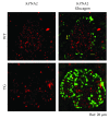
Coimmunostaining of islets for glucagon and KPNA2. Pancreas sections from aged group of mice [WTs (upper panels) and TGs (lower panels)] were costained with antibodies to glucagon (green) and KPNA2 (red) and stained with DAPI (blue) for nuclear staining. Bar, 20 μm. As a whole in the islet, the expression of KPNA2 was much lower in TGs than in WTs. Typical cells with KPNA2 and glucagon double-positive are denoted by yellow triangles in both WTs and TGs. A noticeable tendency exists among cells in the islet: those cells weakly stained with KPNA2, colocalized more in glucagon-positive cells than in non-glucagon-positive cells (upper panels), which suggests that the expression of KPNA2 in α-cells is lower than that in β-cells. This tendency for KPNA2 expression is discernible also in TGs (lower panels). In TGs, glucagon-positive cells were observed throughout the islets.
Figure 7
Figure 7
Islet fibrosis and infiltration of immune cells in the islets of aged group of mice (left panels, WTs; right panels, TGs). Representative cases are shown in each genetic group (in TGs, the islets harboring intra-islet ducts are shown). Bar, 50 μm. (a) Picro-Sirius red staining of the islets. The fibrosis, shown in the red fibrotic structures, is discernible inside the islet of TG. The lumen in the intra-islet duct is also stained with Picro-Sirius red. In the WT case, almost no such structures were observed inside the islet. (b) Immunostaining of islets for F4/80 with alcian blue staining. Representative images are shown in each genetic group. No F4/80-positive cells are observed in the islet of WT. In contrast, several F4/80-positive cells (brown) are discernible near or inside the islets in TG (yellow triangles). The inner side of the component of lumen in the intra-islet duct is stained nonspecifically. (c) Immunostaining of islets for CD3 with alcian blue staining. CD3-positive cells (brown) are recruited to the islets of TG. No CD3-positive cells are observed in the islets of WTs.
Figure 8
Figure 8
mRNA expression of Reg2 and Reg3β in the pancreas. Pancreases were collected from TGs as well as WTs at 4 weeks of age. The relative levels of mRNA were measured by real-time PCR and normalized to the corresponding mHPRT mRNA levels. In each representation, the mean value for WTs was set to 1. Data are means ± SE (WTs, n = 7; TGs, n = 6; P < 0.01 and ∗∗ P < 0.05, t test).

Similar articles

Cited by

References

    1. van der Horst G. T. J., Muijtjens M., Kobayashi K., et al. Mammalian Cry1 and Cry2 are essential for maintenance of circadian rhythms. Nature. 1999;398(6728):627–630. doi: 10.1038/19323. - DOI - PubMed
    1. Kume K., Zylka M. J., Sriram S., et al. mCRY1 and mCRY2 are essential components of the negative limb of the circadian clock feedback loop. Cell. 1999;98(2):193–205. doi: 10.1016/S0092-8674(00)81014-4. - DOI - PubMed
    1. Okano S. Unique aspects of cryptochrome in chronobiology and metabolism, pancreatic β-cell dysfunction, and regeneration: research into cysteine414-alanine mutant CRY1. Journal Diabetes Research. 2016;2016, article 3459246:1–9. doi: 10.1155/2016/3459246. - DOI - PMC - PubMed
    1. Okano S., Akashi M., Hayasaka K., Nakajima O. Unusual circadian locomotor activity and pathophysiology in mutant CRY1 transgenic mice. Neuroscience Letters. 2009;451(3):246–251. doi: 10.1016/j.neulet.2009.01.014. - DOI - PubMed
    1. Okano S., Hayasaka K., Igarashi M., Iwai H., Togashi Y., Nakajima O. Non-obese early onset diabetes mellitus in mutant cryptochrome1 transgenic mice. European Journal of Clinical Investigation. 2010;40(11):1011–1017. doi: 10.1111/j.1365-2362.2010.02359.x. - DOI - PubMed

LinkOut - more resources