

Spatial and Temporal Characteristics of Visual Field Progression in Glaucoma Assessed by Parallel Factor Analysis
- PMID: 31179660
- PMCID: PMC6557789
- DOI: 10.3341/kjo.2019.0004
Spatial and Temporal Characteristics of Visual Field Progression in Glaucoma Assessed by Parallel Factor Analysis
Abstract
Purpose: To explore spatial and temporal characteristics of glaucomatous visual field (VF) progression through multi-way decomposition of data.
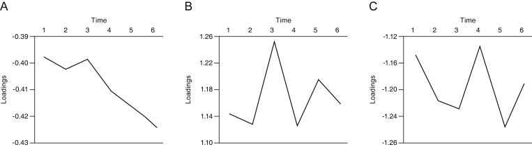
Methods: Six serial VF exams with intervals of 6.0 ± 1.0 months in 121 pre-perimetric glaucoma eyes and 80 perimetric glaucoma eyes were arranged into a three-dimensional cube. The data were decomposed using parallel factor analysis.
Results: Three tri-linear components (i.e., spatial scores, temporal loadings, and subject-specific loadings) were identified. Component 1 clearly showed differences between superior and inferior hemispheres, linear trends over time, and wide variability in perimetric glaucoma. Findings were compatible with well-known characteristics of glaucomatous VF defects. Component 2 showed nasal and central areas in contrast with superior, inferior, and temporal peripheral locations, whereas component 3 showed a contrast between nasal and temporal hemispheres. Both components 2 and 3 failed to show clear temporal trends.
Conclusions: Identification of spatio-temporal patterns shows new possibilities for a multi-way decomposition methodology for earlier diagnosis and prediction of glaucomatous VF progression.
Keywords: Glaucoma; Multi-way decomposition; Parallel factor analysis; Visual fields.
© 2019 The Korean Ophthalmological Society.
Conflict of interest statement
No potential conflict of interest relevant to this article was reported.
Figures


Similar articles
-
Pattern of Macular Ganglion Cell-Inner Plexiform Layer Defect Generated by Spectral-Domain OCT in Glaucoma Patients and Normal Subjects.J Glaucoma. 2015 Oct-Nov;24(8):583-90. doi: 10.1097/IJG.0000000000000231. J Glaucoma. 2015. PMID: 25719232
-
Macular Ganglion Cell Layer Assessment to Detect Glaucomatous Central Visual Field Progression.Korean J Ophthalmol. 2016 Dec;30(6):451-458. doi: 10.3341/kjo.2016.30.6.451. Epub 2016 Dec 6. Korean J Ophthalmol. 2016. PMID: 27980364 Free PMC article.
-
Diagnostic ability of macular ganglion cell asymmetry for glaucoma.Clin Exp Ophthalmol. 2015 Nov;43(8):720-6. doi: 10.1111/ceo.12545. Epub 2015 Jul 1. Clin Exp Ophthalmol. 2015. PMID: 25939316
-
Functional assessment of glaucoma: Uncovering progression.Surv Ophthalmol. 2020 Nov-Dec;65(6):639-661. doi: 10.1016/j.survophthal.2020.04.004. Epub 2020 Apr 26. Surv Ophthalmol. 2020. PMID: 32348798 Free PMC article. Review.
-
Prediction of visual field progression in glaucoma: existing methods and artificial intelligence.Jpn J Ophthalmol. 2023 Sep;67(5):546-559. doi: 10.1007/s10384-023-01009-3. Epub 2023 Aug 4. Jpn J Ophthalmol. 2023. PMID: 37540325 Review.
References
MeSH terms
LinkOut - more resources
Full Text Sources
Medical
