. 2020 May 22;75(6):1021-1030.
doi: 10.1093/gerona/glz105.

Long-Term Intranasal Insulin Aspart: A Profile of Gene Expression, Memory, and Insulin Receptors in Aged F344 Rats

Affiliations

Long-Term Intranasal Insulin Aspart: A Profile of Gene Expression, Memory, and Insulin Receptors in Aged F344 Rats

Hilaree N Frazier et al. J Gerontol A Biol Sci Med Sci. .

Abstract

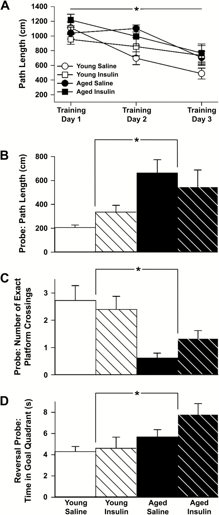
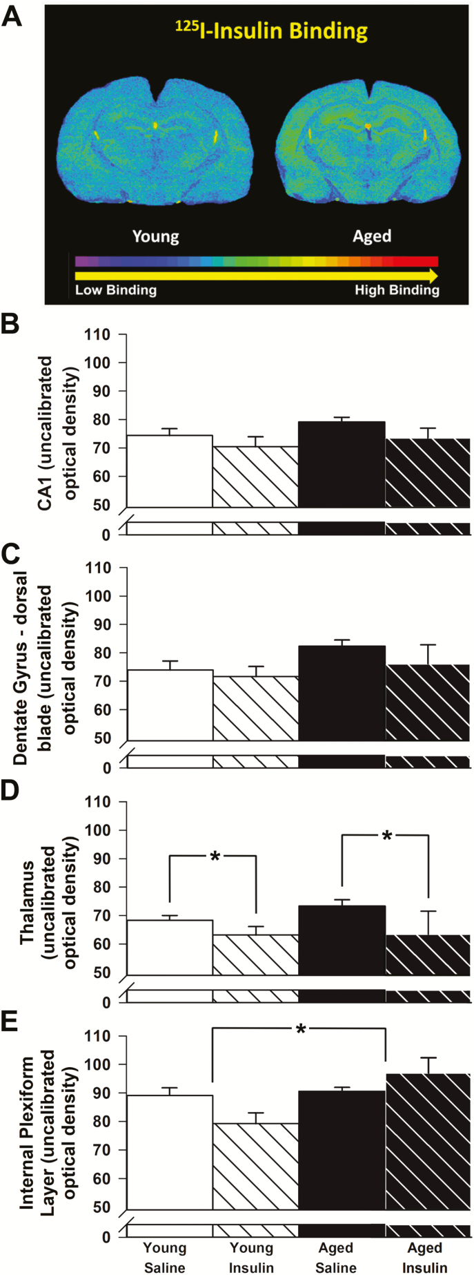
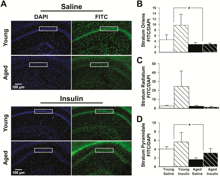
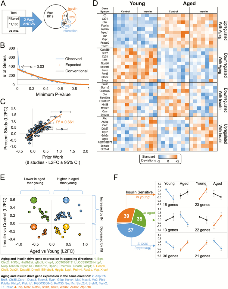
Intranasal insulin is a safe and effective method for ameliorating memory deficits associated with pathological brain aging. However, the impact of different formulations and the duration of treatment on insulin's efficacy and the cellular processes targeted by the treatment remain unclear. Here, we tested whether intranasal insulin aspart, a short-acting insulin formulation, could alleviate memory decline associated with aging and whether long-term treatment affected regulation of insulin receptors and other potential targets. Outcome variables included measures of spatial learning and memory, autoradiography and immunohistochemistry of the insulin receptor, and hippocampal microarray analyses. Aged Fischer 344 rats receiving long-term (3 months) intranasal insulin did not show significant memory enhancement on the Morris water maze task. Autoradiography results showed that long-term treatment reduced insulin binding in the thalamus but not the hippocampus. Results from hippocampal immunofluorescence revealed age-related decreases in insulin immunoreactivity that were partially offset by intranasal administration. Microarray analyses highlighted numerous insulin-sensitive genes, suggesting insulin aspart was able to enter the brain and alter hippocampal RNA expression patterns including those associated with tumor suppression. Our work provides insights into potential mechanisms of intranasal insulin and insulin resistance, and highlights the importance of treatment duration and the brain regions targeted.

Keywords: Animal model; Antiaging; Cognitive decline.


Figures

Figure 1.
Figure 1.
Spatial learning and memory. (A) Path length to goal measures from 43 animals (young saline n = 11, young aspart n = 10, aged saline n = 13, aged aspart n = 9) across 3 days of training showed improved learning over time, though no distinct drug effect was noted. (B) Memory recall on the probe task (24 h) shows young animals (n = 21) identifying the platform location more readily than aged animals (n = 22). (C) During the full 60 s probe task, young animals crossed the exact platform location more often. (D) The 72-h reversal probe showed that the young animals spent significantly more time in the new goal quadrant compared to the aged. Data represent means ± SEM. Asterisks (*) indicate significance at p < 0.05.
Figure 2.
Figure 2.
125I-Insulin receptor binding. (A) Representative images of 125I-insulin receptor binding on a young and aged control brain section. (B) No significant differences with age or insulin treatment were found in field CA1 of the hippocampus (n = 5 per group). (C) Although greater binding of insulin to the dorsal blade of the dentate gyrus was seen with age, this increase was not significant (p = 0.10). (D) Binding in the thalamus decreased significantly with long-term INI. (E) Binding in the internal plexiform layer of the olfactory bulb increased significantly with age. A significant interaction term was also noted, with intranasal insulin (INI) decreasing 125I-insulin binding in young while increasing it in aged. Data represent means ± SEM. Asterisks (*) indicate significance at p < 0.05.
Figure 3.
Figure 3.
Insulin receptor (IR) immunofluorescence. (A) DAPI fluorescence (left) was used to normalize all FITC fluorescence (right) for each hippocampal section quantified. Immunopositive signals representing the presence of the IR were quantified within each region of interest (ROI; white boxes). Equally sized ROIs were used to quantify immunopositive areas across strata oriens (B), radiatum (C), and pyramidale (D) subfields. Strata oriens and pyramidale both showed a significant decrease in IR fluorescence with age (young n = 11, aged n = 15). Data represent means ± SEM. Asterisks (*) indicate significance at p < 0.05.
Figure 4.
Figure 4.
Microarray analyses. (A) Total gene set filtered to remove low-intensity signals yielded 11,160 genes. Two-way analysis of variance (ANOVA) analysis from 26 animals (young saline n = 6, young aspart n = 5, aged saline n = 8, aged aspart n = 7) identified ~1,500 genes that were significant by main effects of age, insulin, and/or the interaction (B) p value frequency histogram shows the increase in the number of significant genes with α < 0.03. The conventional line (gray) delineates a cutoff for significance near 112 as the first percentile (p < 0.01) of the 11,160 filtered genes. The orange line represents the p values obtain when testing for significance across a set of 11,160 randomly generated numbers through a two-way analysis of variance (ANOVA). The blue line highlights the p values obtain from our data set. (C) We validated ~1,000 age-sensitive genes across our prior studies and found a significant correlation with prior work. (D) Heatmap of significant genes (top 10) separated across participants by aging or drug effects. Each column represents one animal and each row represents one gene. Color-coded signal intensity values (standardized: orange represents increase, blue represents decrease) are shown. (E) Genes significant by both main effects (63) fall into four categories: 2 in the same direction (quadrants 4 and 2), and 2 in the opposite direction (quadrants 1 and 3). (F) Genes within the significant interaction space (~130) are divided as those modified in young animals (top), in aged animals (middle), or in both (bottom). This result suggests insulin sensitivity in the brain may not differ across aging (39 genes changed in young, and 35 changed in aged). Data represent means ± SEM.

Similar articles

Cited by

References

    1. Lochhead JJ, Thorne RG. Intranasal delivery of biologics to the central nervous system. Adv Drug Deliv Rev. 2012;64:614–628. doi:10.1016/j.addr.2011.11.002 - PubMed
    1. Benedict C, Hallschmid M, Schmitz K, et al. . Intranasal insulin improves memory in humans: superiority of insulin aspart. Neuropsychopharmacology. 2007;32:239–243. doi:10.1038/sj.npp.1301193 - PubMed
    1. Brabazon F, Wilson CM, Jaiswal S, Reed J, Frey WH, Byrnes KR. Intranasal insulin treatment of an experimental model of moderate traumatic brain injury. J Cereb Blood Flow Metab. 2017;37:3203–3218. doi:10.1177/0271678X16685106 - PMC - PubMed
    1. Born J, Lange T, Kern W, McGregor GP, Bickel U, Fehm HL. Sniffing neuropeptides: a transnasal approach to the human brain. Nat Neurosci. 2002;5:514–516. doi:10.1038/nn849 - PubMed
    1. Thorne RG, Emory CR, Ala TA, Frey WH II. Quantitative analysis of the olfactory pathway for drug delivery to the brain. Brain Res. 1995;692:278–282. doi:10.1016/0006-8993(95)00637-6 - PubMed

Publication types