. 2019 Jun 10;14(6):e0216979.
doi: 10.1371/journal.pone.0216979. eCollection 2019.

Crystal structures of Moorella thermoacetica cyanuric acid hydrolase reveal conformational flexibility and asymmetry important for catalysis

Affiliations

Crystal structures of Moorella thermoacetica cyanuric acid hydrolase reveal conformational flexibility and asymmetry important for catalysis

Ke Shi et al. PLoS One. .

Abstract

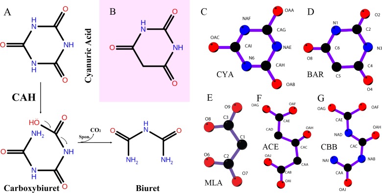
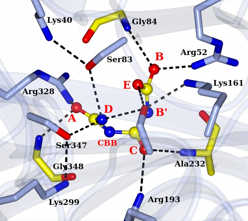
An ancient enzyme family responsible for the catabolism of the prebiotic chemical cyanuric acid (1,3,5-triazine-2,4,6-triol) was recently discovered and is undergoing proliferation in the modern world due to industrial synthesis and dissemination of 1,3,5-triazine compounds. Cyanuric acid has a highly stabilized ring system such that bacteria require a unique enzyme with a novel fold and subtle active site construction to open the ring. Each cyanuric acid hydrolase monomer consists of three isostructural domains that coordinate and activate the three-fold symmetric substrate cyanuric acid for ring opening. We have now solved a series of X-ray structures of an engineered, thermostable cyanuric acid ring-opening enzyme at 1.51 ~ 2.25 Å resolution, including various complexes with the substrate, a tight-binding inhibitor, or an analog of the reaction intermediate. These structures reveal asymmetric interactions between the enzyme and bound ligands, a metal ion binding coupled to conformational changes and substrate binding important for enzyme stability, and distinct roles of the isostructural domains of the enzyme. The multiple conformations of the enzyme observed across a series of structures and corroborating biochemical data suggest importance of the structural dynamics in facilitating the substrate entry and the ring-opening reaction, catalyzed by a conserved Ser-Lys dyad.


Conflict of interest statement

I have read the journal's policy and the authors of this manuscript have the following competing interests: Dr. Lawrence Wackett owns equity in and is entitled to royalties from Minnepura Technologies, Inc., a company involved in the development, commercialization and marketing of patented encapsulated biological platforms for water treatment. The University of Minnesota also has equity and royalty interest in Minnepura. These interests have been reviewed and managed by the University of Minnesota in accordance with its conflict of interest policies. The other authors have no conflict of interest to declare. This does not alter our adherence to PLOS ONE policies on sharing data and materials.

Figures

Fig 1
Fig 1
A. Schematic representation of the ring opening reaction catalyzed by Cyanuric Acid Hydrolase (CAH). B. Barbituric acid, differing from cyanuric acid by only one non-hydrogen atom, inhibits the CAH activity. C-G. The chemicals discussed in the paper, either used for soaking, or in the crystallization solution or enzymatic reaction intermediates. C. Cyanuric acid, substrate of the enzyme. D. Barbituric acid, a tight binding iso-structural inhibitor of the enzyme. E. Malonate, one of the components used in crystallization, a mimic of the final product of the enzymatic reaction, biuret. F. 3-Oxopentanedioic acid, or 1,3-Acetonedicarboxylic acid, a mimic of the open-ring intermediate, designed for soaking to form enzyme-intermediate complex. G. The theoretical open-ring intermediate generated by the enzymatic reaction.
Fig 2
Fig 2. Domain architecture of cyanuric acid hydrolase.
A. RMCAH monomer shown in a ribbon diagram viewed along the pseudo three-fold axis and with the three domains colored differently: Domain 1 (residues 1–102) is in green, Domain 2 (111–247) in blue and Domain 3 (253–367) in magenta. Malonate observed at the domain interface is shown in a ball-and-stick representation. The 2mFo-DFc electron density map contoured at 1.0 σ level is overlaid and shown in orange-colored mesh. B. A superposition of the three domains color-coded as in A with the conserved Ser-Lys dyads and the partner Arg side chains shown in sticks. The arginine is located in the middle of the longest helix of each domain, while the serine and lysine are from the 3rd and 2nd β-strands of the 4-β-pleated core, respectively. The three corresponding serines superpose very well, so do the three lysines and arginines. C. Same as B with a view rotated ~90° about the vertical axis.
Fig 3
Fig 3. Architecture and properties of the RMCAH tetramer.
A. Domain 3 and domain 1 form the tetramer core. An extended loop from the domain 3 forms extensive inter-monomer interactions. B. The detailed polar interactions made by the extended loop of domain 3 in the tetramer core. C. Domains 2 of monomer M4 depicted in purple and that of M2 depicted in green, showing their lack of inter-monomer interactions in the tetramer. D. M2 and M4 are shown in B-factor scaled worm. The color with increased B-factor is depicted in blue
Fig 4
Fig 4. Positioning of MLA in the RMCAH active site.
A. Top-view of MLA in the active site overlaid with 2mFo-DFc electron density map contoured at 1.0 σ level, depicted in blue mesh. A barbituric acid (BAR) molecule from RMCAH/BAR structure (depicted in green lines, will be discussed in detail later) is depicted as a reference to show the substrate plane and demonstrate the relative location of MLA atoms. The three carbonyl oxygen positions of BAR are designated A, B, and C for simplifying structural descriptions. The three intervening atoms corresponding to amide nitrogen atoms in CYA are named A’, B’, and C’. B. Side-view of interactions between malonate and RMCAH active site residues.
Fig 5
Fig 5. Comparison of the open and closed conformations of RMCAH, and the relationship between Ca2+ ion binding and RMCAH conformation.
A. Molecular surface of the RMCAH/MLA complex, colored according to electrostatic potential. Two channels differ greatly in their charge properties. B. Top view of superposition of the APO open conformation (green) with the MLA-bound closed conformation (magenta). C. Side view of the superposition, with the domain 3 in front and facing to the right, showing displacement of the domain 2. See also S1 Video. D. 2mFo-DFc simulated annealing composite omit map showing the calcium binding loop contoured at 1.5σ level. The same map for Ca2+ is shown in orange color. The anomalous difference Fourier map for Ca2+ is shown in pink and contoured at 3σ level. E. A close-up view of the interactions made by the short segment Ser347-Gly348-Gly349-Ala350-Glu351-His352-Gln353 in domain 3 in the closed conformation (magenta). The backbone trace for the open conformation is shown in green for comparison.
Fig 6
Fig 6. RMCAH with CYA bound.
A. 2mFo-DFc simulated annealing composite omit electron density map contoured at 1.0 σ level depicted in blue mesh, and mFo-DFc omit map with only MLA modeled in contoured at 3.0 σ level depicted in green mesh. As the strong residual density shows that there should be at least partial occupancy of CYA in the active site of the monomer M2, a CYA is placed in the map to show its existence. B. Cut-away surface representation showing two channels to the active site, indicated by green and blue arrows. One cyanuric acid molecule binds at the entry of the 1st channel. C. Interactions formed between CYA and two protein monomers at the entry of the ingress channel. D. A string of water molecules lined up in the ingress channel. The size of the substrate can be seen larger than the diameter of the channel at the narrowest point. All maps in B, C, D are the mFo-DFc simulated annealing composite omit map shown in pink or blue mesh.
Fig 7
Fig 7. RMCAH with 1,3-acetonedicarboxylic acid (ACE) bound.
A. Top-view of 2mFo-DFc electron density map contoured at 1.0 σ level, depicted in blue mesh on ACE. ACE is shown in ball-and-stick. Hydrogen bonds are indicated by the black dotted lines. A reference barbituric acid molecule from the RMCAH/BAR structure (depicted in green lines) is overlaid to show the substrate plain and the relative location of ACE atoms. B. Side-view of ACE bound in the active site.
Fig 8
Fig 8. RMCAH with barbituric acid (BAR) bound and the detailed environment for each of the three carbonyl oxygen atoms.
A. 2mFo-DFc electron density map contoured at 1.5σ level, depicted in blue mesh. B. Site A, the αFL for Ser83 and Ser347 are shown in red arrows. The hydrogen bonding interactions for the carbonyl group are shown by dashed lines. C. Site B. Only Ser231 is close enough for initiating the reaction. The distance/αBD/αFL of Ser231 to the carbonyl group carbon of BAR are 3.4Å/110°/5°. Side chain of Glu235 forms one hydrogen bond with the side chain of Arg52. D. Site C. the distance/αBD/αFL of Ser231 to the carbonyl group carbon of BAR are 3.3Å/106°/7°. Side chain of Arg193 forms two hydrogen bonding interactions with side chain of Glu351, which is in the Ca2+ coordination loop.
Fig 9
Fig 9. Hydrogen-deuterium exchange mass-spectroscopy (HDX-MS).
A. Heat map showing the relative fractional deuterium uptake at indicated time points for proteolytic fragments of apo RMCAH (DMSO control, lower strip) and RMCAH/BAR complex (upper strip). Lowest exchange rate is depicted in blue and highest in red. Residues not detected in MS are shown in white. B, C. Tube representation of the difference in HDX rate between RMCAH and RMCAH/BAR, mapped on the closed BAR-bound (B) and apo (C) structures of RMCAH. Smaller differences are depicted in thin tube and blue color, while larger differences are in thick tube and red color. Note that the color-coding in (B) and (C) has a different meaning from that in (A).
Fig 10
Fig 10. Atomic model for the reaction intermediate, carboxybiuret, bound in the active site of RMCAH.
The model is based on the crystal structure of the RMCAH/ACE complex shown in Fig 7 and the active site properties revealed by other RMCAH/ligand structures.

References

    1. Huthmacher KM, D. Cyanuric acid and cyanuric chloride. Ullmann's Encyclopedia of Industrial Chemistry. 2000;11.
    1. Hayatsu R, Studier M. H., Oda A., Fuse K., Anders E. Origin of organic matter in early solar system. II. 1968;32:175–90. 10.1016/S0016-7037(68)80003-1 - DOI
    1. Hatfield SE. Applications of triazine chemistry: education, remediation, and drug delivery. TX: Texas A&M University, College Station; 2007.
    1. Saldick J. Biodegradation of cyanuric acid. Applied microbiology. 1974;28(6):1004–8. Epub 1974/12/01. - PMC - PubMed
    1. Seffernick JL, Shapir N, Schoeb M, Johnson G, Sadowsky MJ, Wackett LP. Enzymatic degradation of chlorodiamino-s-triazine. Applied and environmental microbiology. 2002;68(9):4672–5. Epub 2002/08/30. 10.1128/AEM.68.9.4672-4675.2002 - DOI - PMC - PubMed

Publication types

LinkOut - more resources