. 2019 Jun 10;14(6):e0218130.
doi: 10.1371/journal.pone.0218130. eCollection 2019.

L-dopa response pattern in a rat model of mild striatonigral degeneration

Affiliations

L-dopa response pattern in a rat model of mild striatonigral degeneration

Christine Kaindlstorfer et al. PLoS One. .

Abstract

Background: Unresponsiveness to dopaminergic therapies is a key feature in the diagnosis of multiple system atrophy (MSA) and a major unmet need in the treatment of MSA patients caused by combined striatonigral degeneration (SND). Transgenic, alpha-synuclein animal models do not recapitulate this lack of levodopa responsiveness. In order to preclinically study interventions including striatal cell grafts, models that feature SND are required. Most of the previous studies focused on extensive nigral and striatal lesions corresponding to advanced MSA-P/SND. The aim of the current study was to replicate mild stage MSA-P/SND with L-dopa failure.

Methods and results: Two different striatal quinolinic acid (QA) lesions following a striatal 6-OHDA lesion replicating mild and severe MSA-P/SND, respectively, were investigated and compared to 6-OHDA lesioned animals. After the initial 6-OHDA lesion there was a significant improvement of motor performance after dopaminergic stimulation in the cylinder and stepping test (p<0.001). Response to L-dopa treatment declined in both MSA-P/SND groups reflecting striatal damage of lateral motor areas in contrast to the 6-OHDA only lesioned animals (p<0.01). The remaining striatal volume correlated strongly with contralateral apomorphine induced rotation behaviour and contralateral paw use during L-dopa treatment in cylinder and stepping test (p<0.001).

Conclusion: Our novel L-dopa response data suggest that L-dopa failure can be induced by restricted lateral striatal lesions combined with dopaminergic denervation. We propose that this sequential striatal double-lesion model replicates a mild stage of MSA-P/SND and is suitable to address neuro-regenerative therapies aimed at restoring dopaminergic responsiveness.


Conflict of interest statement

The authors have declared that no competing interests exist.

Figures
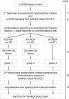
Fig 1
Fig 1. Experimental design.
All 54 male Wistar rats underwent 6-hydroxydopamine (6-OHDA) lesion surgery. The first behavioural assessment included amphetamine induced rotation, cylinder and stepping test with saline (S1) or L-dopa (LD1) treatment. Animals were then randomized into the three experimental groups according to rotation behaviour and L-dopa response. Groups 1 and 2 received the additional quinolinic acid (QA) lesions: group 1: 6-OHDA+severe QA and group 2: 6-OHDA+mild QA, while group 3 served as the Parkinson´s disease control group with the 6-OHDA lesion only. Afterwards, the second behavioural assessment including cylinder and stepping test with saline (S2) or L-dopa treatment (LD2), amphetamine and apomorphine induced rotation was performed. Finally, animals were sacrificed for histological purposes.
Fig 2
Fig 2
L-dopa response patterns (A-F) and related histology (G-H). Data are illustrated as mean ± standard error. L-dopa responsiveness was evaluated by cylinder and stepping test. All animals received the initial 6-hydroxydopamine (6-OHDA) lesion prior to the first behavioural assessment, while only groups 1 and 2 received the additional quinolinic acid (QA) lesions before the second behavioural assessment: group 1: 6-OHDA+severe QA; group 2: 6-OHDA+mild QA; group 3: 6-OHDA. At the first behavioural assessment (A,C,E), all experimental groups showed a significant motor improvement after L-dopa administration compared to saline treatment in contralateral paw use of cylinder test (A), and limb asymmetry score (LAS) of forehand (C) and backhand stepping test (E; p<0.05). At the second behavioural assessment (B,D,F), the L-dopa treatment effect was still significant in cylinder and stepping test, but declined comparably in both MSA-P/SND groups so that only the group 3 showed a sustained motor improvement during dopaminergic stimulation in both motor tests (p<0.05). G-H: The 6-OHDA lesion resulted in a significant reduction of tyrosine hydroxylase positive (TH+) neurons in the ipsilateral substantia nigra pars compacta (SNc) compared to the contralateral SNc (p<0.001; G). Ipsilateral striatal volume was significantly reduced compared to the contralateral side in all groups (***p<0.001) and the QA lesion resulted in a significant reduction of ipsilateral striatal volumes in groups 1 and 2 compared to the 6-OHDA only lesioned group 3 (§§§p<0.001; H). Abbreviations: S1…saline treatment at the first behavioural assessment, LD1…L-dopa treatment at the first behavioural assessment; S2… saline treatment at the second behavioural assessment, LD2…L-dopa treatment at the second behavioural assessment; LAS…limb asymmetry score.
Fig 3
Fig 3. Representative examples of the striatal pathology.
Dopamine- and adenosine 3,5-monophosphate regulated phosphoprotein (DARPP32) immunohistochemistry was used to visualize the striatal projection neurons. Three sections in the rostral, middle and caudal section of the striatum are shown per group.

Similar articles

Cited by

References

    1. Köllensperger M, Geser F, Ndayisaba JP, Boesch S, Seppi K, Ostergaard K, et al. EMSA-SG. Presentation, diagnosis, and management of multiple system atrophy in Europe: final analysis of the European multiple system atrophy registry. Mov Disord. 2010. November 15;25(15):2604–12. 10.1002/mds.23192 - DOI - PubMed
    1. Krismer F, Wenning GK. Multiple system atrophy: insights into a rare and debilitating movement disorder. Nat Rev Neurol. 2017. Apr;13(4):232–243. 10.1038/nrneurol.2017.26 - DOI - PubMed
    1. Ben-Shlomo Y, Wenning GK, Tison F, Quinn NP. Survival of patients with pathologically proven multiple system atrophy: a meta-analysis. Neurology. 1997. February;48(2):384–93. 10.1212/wnl.48.2.384 - DOI - PubMed
    1. Wüllner U, Schmitz-Hübsch T, Abele M, Antony G, Bauer P, Eggert K. Features of probable multiple system atrophy patients identified among 4770 patients with parkinsonism enrolled in the multicentre registry of the German Competence Network on Parkinson's disease. J Neural Transm (Vienna). 2007. September;114(9):1161–5. Epub 2007 May 18. - PubMed
    1. Wenning GK, Geser F, Krismer F, Seppi K, Duerr S, Boesch S, et al. European Multiple System Atrophy Study Group. The natural history of multiple system atrophy: a prospective European cohort study. Lancet Neurol. 2013. March;12(3):264–74. 10.1016/S1474-4422(12)70327-7 - DOI - PMC - PubMed

Publication types