

Differential effects of antipsychotic drugs on contrast response functions of retinal ganglion cells in wild-type Sprague-Dawley rats and P23H retinitis pigmentosa rats
- PMID: 31181134
- PMCID: PMC6557501
- DOI: 10.1371/journal.pone.0218200
Differential effects of antipsychotic drugs on contrast response functions of retinal ganglion cells in wild-type Sprague-Dawley rats and P23H retinitis pigmentosa rats
Abstract
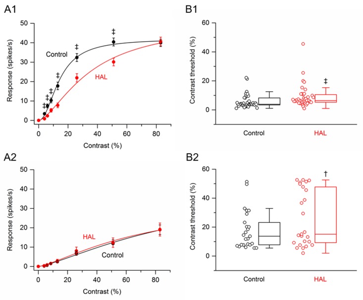
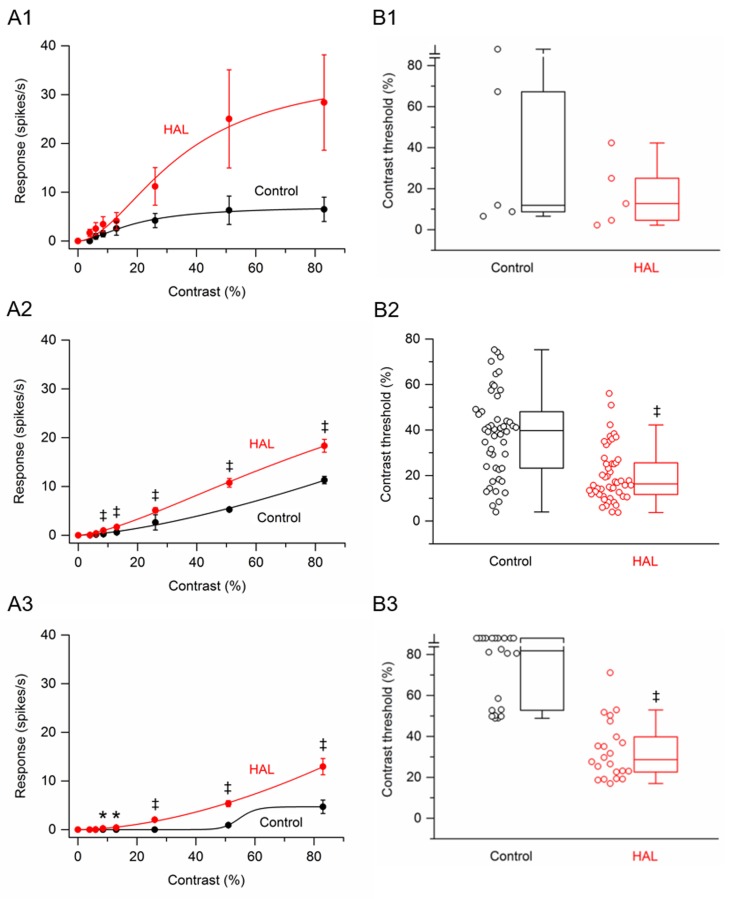
Antipsychotic drugs haloperidol and clozapine have been reported to increase the sensitivity of retinal ganglion cells (RGCs) to flashes of light in the P23H rat model of retinitis pigmentosa. In order to better understand the effects of these antipsychotic drugs on the visual responses of P23H rat RGCs, I examined the responses of RGCs to a drifting sinusoidal grating of various contrasts. In-vitro multielectrode array recordings were made from P23H rat RGCs and healthy Sprague-Dawley (SD) rat RGCs. Retinas were stimulated with a drifting sinusoidal grating with eight values of contrast (0, 4, 6, 8.5, 13, 26, 51, and 83%). Contrast response functions based on response amplitudes were fitted with a hyperbolic ratio function and contrast thresholds were determined from the fitted curves. SD rat RGCs were divided into two categories, saturating and non-saturating cells, based on whether they showed saturation of responses at high contrast levels. Most SD rat RGCs (58%) were saturating cells. Haloperidol and clozapine decreased the responses of saturating SD rat RGCs to all grating contrasts, except for the highest contrast tested. Clozapine also decreased the responses of non-saturating SD rat RGCs to all grating contrasts, except for the highest contrast tested. Haloperidol did not however significantly affect the responses of non-saturating SD rat RGCs. Haloperidol and clozapine increased the contrast thresholds of both saturating and non-saturating cells in SD rat retinas. Most (73%) P23H rat RGCs could be categorized as either saturating or non-saturating cells. The remaining 'uncategorized' cells were poorly responsive to the drifting grating and were analyzed separately. Haloperidol and clozapine increased the responses of non-saturating and uncategorized P23H rat RGCs to most grating contrasts, including the highest contrast tested. Haloperidol and clozapine also increased the responses of saturating P23H rat RGCs to most grating contrasts but these increases were not statistically significant. Haloperidol and clozapine decreased the contrast thresholds of saturating cells, non-saturating cells and uncategorized cells in P23H rat retinas, although the decrease in contrast thresholds of saturating cells was not found to be statistically significant. Overall, the findings show that haloperidol and clozapine have differential effects on the contrast response functions of SD and P23H rat RGCs. In contrast to the effects observed on SD rat RGCs, both haloperidol and clozapine increased the responsiveness of P23H rat RGCs to both low and high contrast visual stimuli and decreased contrast thresholds.
Conflict of interest statement
The authors have declared that no competing interests exist.
Figures


Similar articles
-
Effects of GABACR and mGluR1 antagonists on contrast response functions of Sprague-Dawley and P23H rat retinal ganglion cells.PLoS One. 2017 Dec 18;12(12):e0189980. doi: 10.1371/journal.pone.0189980. eCollection 2017. PLoS One. 2017. PMID: 29253887 Free PMC article.
-
Effects of Antipsychotic Drugs Haloperidol and Clozapine on Visual Responses of Retinal Ganglion Cells in a Rat Model of Retinitis Pigmentosa.J Ocul Pharmacol Ther. 2016 Dec;32(10):685-690. doi: 10.1089/jop.2016.0102. Epub 2016 Oct 27. J Ocul Pharmacol Ther. 2016. PMID: 27788033
-
Activation of ganglion cells in wild-type and P23H rat retinas with a small subretinal electrode.Exp Eye Res. 2012 Jun;99:71-7. doi: 10.1016/j.exer.2012.03.016. Epub 2012 Apr 20. Exp Eye Res. 2012. PMID: 22542904
-
Blocking GABA(C) receptors increases light responsiveness of retinal ganglion cells in a rat model of retinitis pigmentosa.Exp Eye Res. 2012 Dec;105:21-6. doi: 10.1016/j.exer.2012.10.005. Epub 2012 Oct 16. Exp Eye Res. 2012. PMID: 23085337
-
Effects of Dopamine D2-Like Receptor Antagonists on Light Responses of Ganglion Cells in Wild-Type and P23H Rat Retinas.PLoS One. 2015 Dec 30;10(12):e0146154. doi: 10.1371/journal.pone.0146154. eCollection 2015. PLoS One. 2015. PMID: 26717015 Free PMC article.
Cited by
-
Neuroprotection of photoreceptors by combined inhibition of both Fas and autophagy pathways in P23H mice.Cell Death Dis. 2025 Jul 1;16(1):469. doi: 10.1038/s41419-025-07793-9. Cell Death Dis. 2025. PMID: 40592891 Free PMC article.
-
Loss of Fas Receptor Function Preserves Photoreceptor Structure and Function in Two Mouse Models of Inherited Retinal Degeneration.Invest Ophthalmol Vis Sci. 2022 Sep 1;63(10):5. doi: 10.1167/iovs.63.10.5. Invest Ophthalmol Vis Sci. 2022. PMID: 36083588 Free PMC article.
-
Loss of αA or αB-Crystallin Accelerates Photoreceptor Cell Death in a Mouse Model of P23H Autosomal Dominant Retinitis Pigmentosa.Int J Mol Sci. 2021 Dec 22;23(1):70. doi: 10.3390/ijms23010070. Int J Mol Sci. 2021. PMID: 35008496 Free PMC article.
-
Preservation of retinal structure and function in two mouse models of inherited retinal degeneration by ONL1204, an inhibitor of the Fas receptor.Cell Death Dis. 2024 Aug 8;15(8):576. doi: 10.1038/s41419-024-06970-6. Cell Death Dis. 2024. PMID: 39117629 Free PMC article.
References
-
- Sacchetti M, Mantelli F, Rocco ML, Micera A, Brandolini L, Focareta L, et al. Recombinant human nerve growth factor treatment promotes photoreceptor survival in the retinas of rats with retinitis pigmentosa. Cur. Eye Res. 2017; 42: 1064–1068. - PubMed
Publication types
MeSH terms
Substances
Grants and funding
LinkOut - more resources
Full Text Sources
