. 2019 Jun 7;19(11):2595.
doi: 10.3390/s19112595.

Optical Fiber Gratings Immunoassays

Affiliations

Optical Fiber Gratings Immunoassays

Médéric Loyez et al. Sensors (Basel). .

Abstract

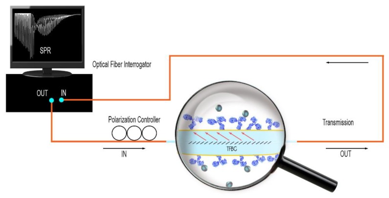
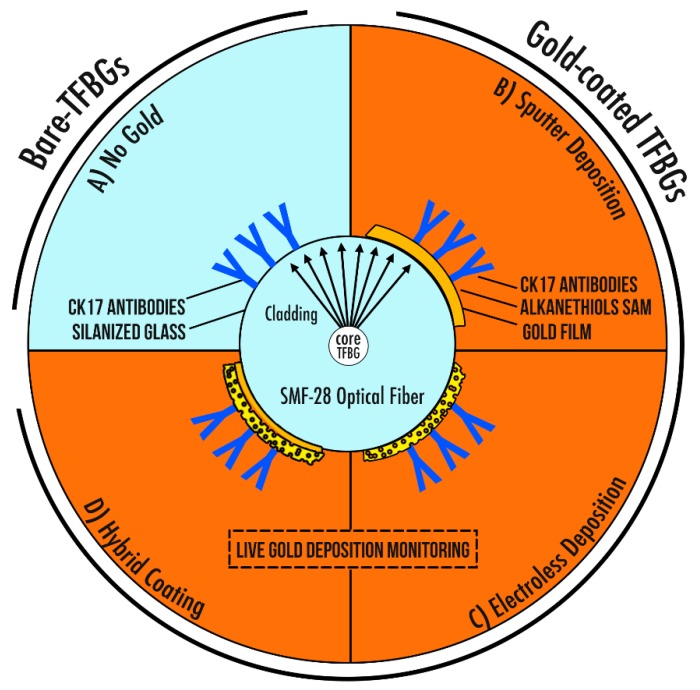
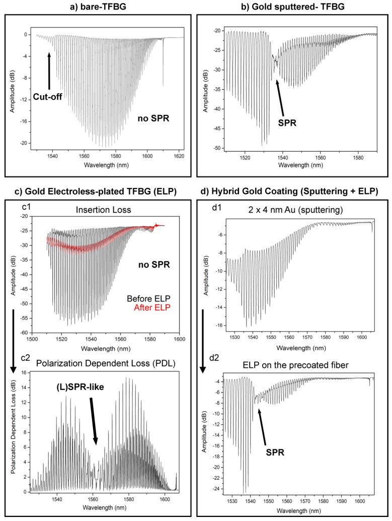
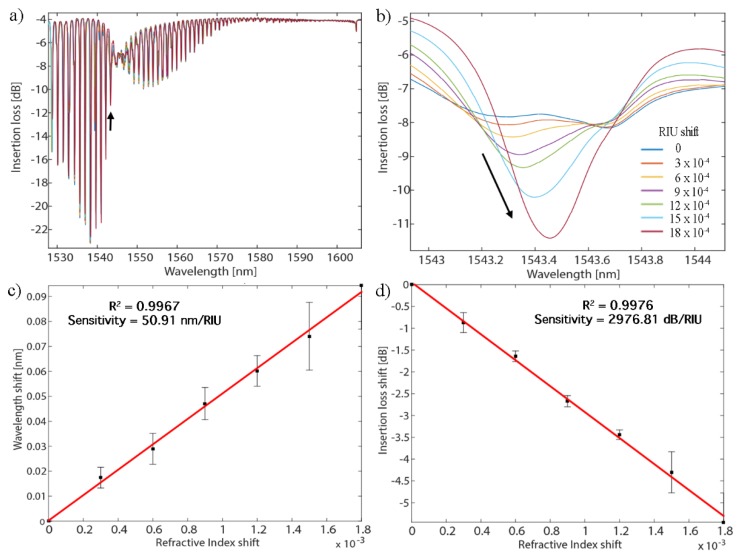
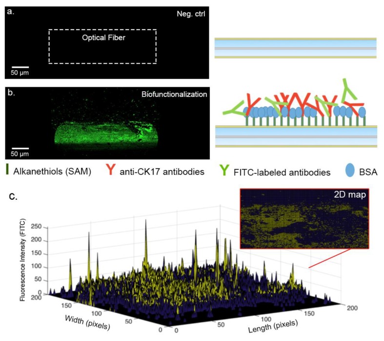
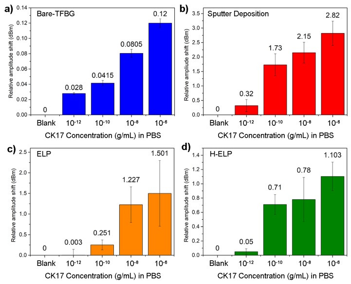
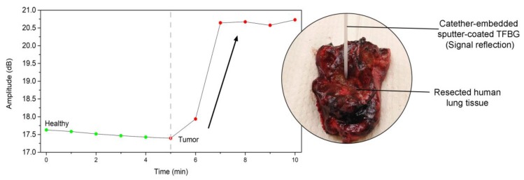
Optical fibers are of growing interest for biosensing, especially for point-of-care and biomedical assays. Their intrinsic properties bestow them sought-after assets for the detection of low concentrations of analytes. Tilted fiber Bragg gratings (TFBGs) photo-inscribed in the core of telecommunication-grade optical fibers are known to be highly-sensitive refractometers. In this work, we present different strategies to use them for label-free immunoassays. Bare, gold-sputtered, gold-electroless-plated (ELP) and hybrid configurations are biofunctionalized with antibodies, aiming at the detection of cancer biomarkers. We discuss the relative performances of the tested configurations and show that each leads to singular key features, which therefore drives their selection as a function of the target application. The most sensitive configuration presents a limit of detection of 10-12 g/mL in laboratory settings and was successfully used ex vivo in freshly resected lung tissues.

Keywords: biomarker; biosensing; immunoassays; optical fibers; tilted fiber bragg gratings.


Conflict of interest statement

The authors declare no conflict of interest

Figures

Figure 1
Figure 1
Scheme of the experimental setup in transmission. The gold-sputtered tilted fiber Bragg grating (TFBG) is connected using pigtails (SMF-28) to a polarization controller and an optical interrogator.
Figure 2
Figure 2
Scheme of the four studied sensing configurations. (A) The bare-TFBGs with adsorbed antibodies on the silica surface. (B) The sputter deposition under vacuum and the covalent bonding of anti-CK17 antibodies on the gold film. (C) The electroless deposition of gold and the covalent bonding of anti-CK17 antibodies on the agglomerated Au particles. (D) A hybrid-coating coupling a deposition of 4 nm Au by sputter-coater followed by an ELP deposition, which is functionalized using covalent bonding of anti-CK17 antibodies.
Figure 3
Figure 3
(a) Bare-TFBG amplitude spectrum monitored in PBS with the sensing part located at the cut-off area. (b) P-polarized spectrum of a sputter-coated TFBG with the most sensitive modes located at lower neighbor wavelengths of the SPR attenuation. (c1) Transmitted amplitude spectrum before and after ELP process. (c2) The same plating leads to a pronounced change in the PDL spectrum. (d1) The deposition of a ~4 nm-thick gold layer by sputtering does not lead to SPR attenuation. (d2) While the ELP is performed on that layer, the reaction can be stopped at the optimum point to reach SPR excitation using classical insertion loss interrogation.
Figure 4
Figure 4
(a) SEM picture of a sputter-coated optical fiber surface (1 × 50 nm Au). (b) SEM picture of an electroless-plated (ELP) optical fiber surface.
Figure 5
Figure 5
(a) P-polarized spectral evolution of a hybrid gold-coated TFBG configuration immersed in different LiCl solutions. (b) Zoom on the most sensitive mode. (c) Wavelength evolution of that mode as function of RI change (d) Amplitude evolution of that mode as function of refractive index (RI) change for three tested fibers.
Figure 6
Figure 6
Confocal microscopy analysis performed using fluorescein isothiocyanate (FITC)-labeled antibodies. (a) Scans performed with a gold-coated SMF28 optical fiber (no fluorescent antibodies). (b) Scan performed with a gold-coated optical fiber covered with the FITC-labeled antibodies and Bovine Serum Albumine (BSA)-blocked. (c) 3D map obtained by the fluorescence intensity, bringing approximations of the antibodies covered areas.
Figure 7
Figure 7
Cytokeratin-17 detection in Phosphate Buffer Saline (PBS). (a) Detection using the sensitivity of the cut-off area of bare-TFBGs. (b) Detection through the SPR-related modes of gold sputter-coated TFBGs. (c) Detection through the sensitive modes of the polarization dependent loss (PDL) spectra of gold-ELP-TFBGs. (d) Detection through the hybrid configuration. Data show means of amplitude shifts ± standard deviations for three tested fibers, in each condition.
Figure 8
Figure 8
Graph showing the biosensor response in both healthy and tumorous parts of a resected human lung tissue. The biosensor was used at the tip of a catheter.

Similar articles

Cited by

References

    1. Yin M., Gu B., An Q., Yang C., Guan Y.L., Yong K. Recent development of fiber-optic chemical sensors and biosensors: Mechanisms, materials, micro/nano-fabrications and applications. Coord. Chem. Rev. 2018;376:348–392. doi: 10.1016/j.ccr.2018.08.001. - DOI
    1. Gupta B.D., Kant R. Recent advances in surface plasmon resonance based fiber optic chemical and biosensors utilizing bulk and nanostructures. Opt. Laser Technol. 2018;101:144–161. doi: 10.1016/j.optlastec.2017.11.015. - DOI
    1. Shan D., Gerhard E., Zhang C., Tierney J.W., Xie D., Liu Z., Yang J. Polymeric biomaterials for biophotonic applications. Bioact. Mater. 2018;3:434–445. doi: 10.1016/j.bioactmat.2018.07.001. - DOI - PMC - PubMed
    1. Daems D., Pfeifer W., Rutten I., Sacca B., Spasic D., Lammertyn J. 3D DNA origami as programmable anchoring points for bioreceptors in fiber optic surface plasmon resonance biosensing. ACS Appl. Mater. Interfaces. 2018;10:23539–23547. doi: 10.1021/acsami.8b04757. - DOI - PubMed
    1. Pollet J., Delport F., Janssen K.P.F., Jans K., Maes G., Pfeiffer H., Wevers M., Lammertyn J. Fiber optic SPR biosensing of DNA hybridization and DNA-protein interactions. Biosens. Bioelectron. 2009;25:864–869. doi: 10.1016/j.bios.2009.08.045. - DOI - PubMed

Substances

LinkOut - more resources