

Comparison of Different Polarization Sensitive Second Harmonic Generation Imaging Techniques
- PMID: 31181703
- PMCID: PMC6632172
- DOI: 10.3390/mps2020049
Comparison of Different Polarization Sensitive Second Harmonic Generation Imaging Techniques
Abstract
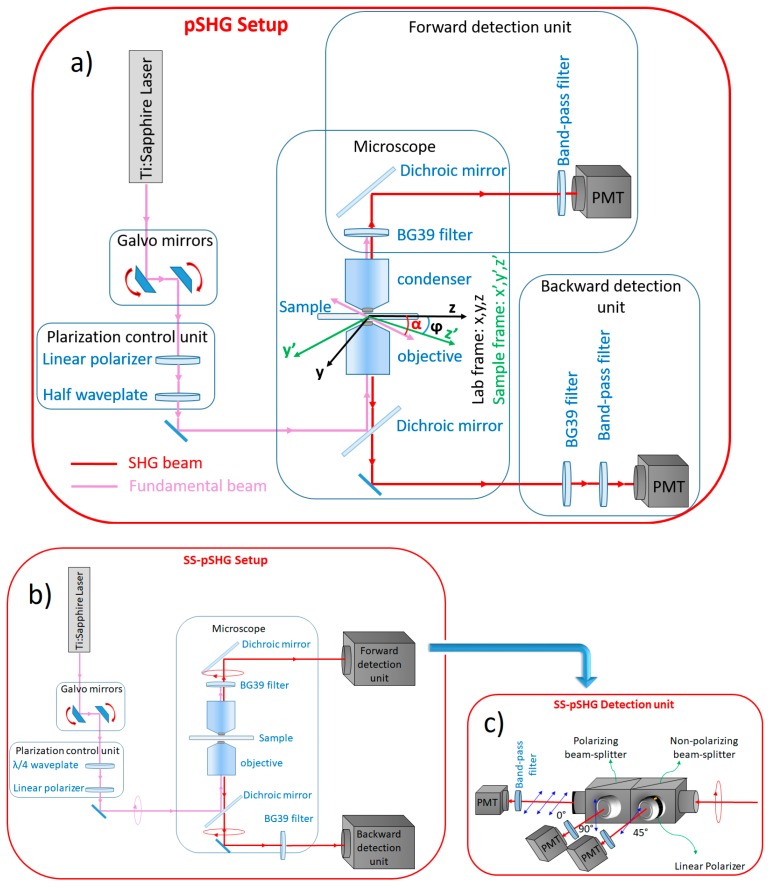
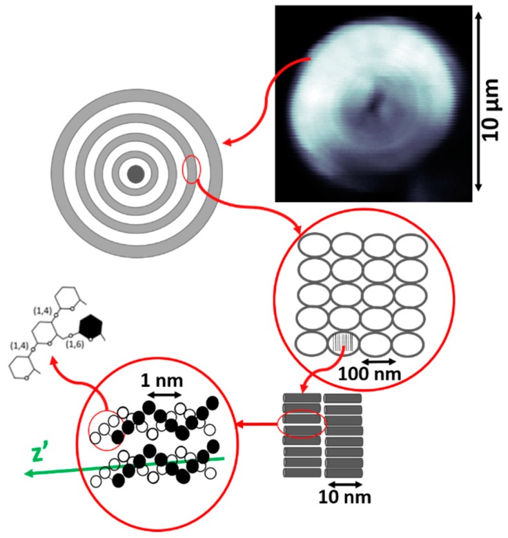
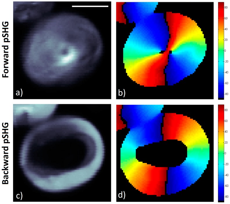
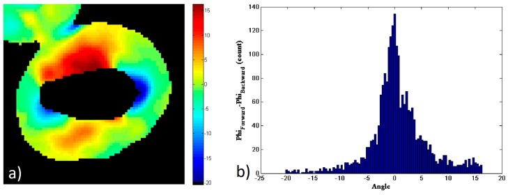
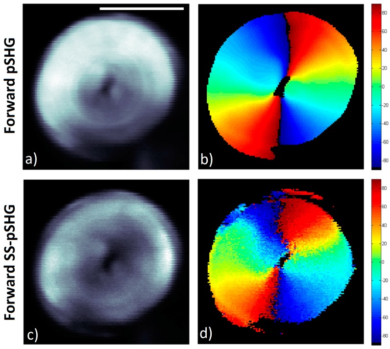
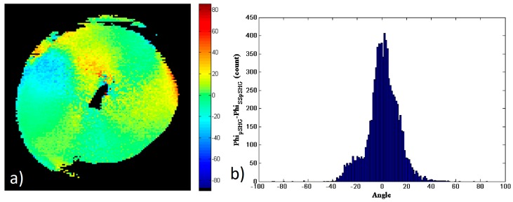
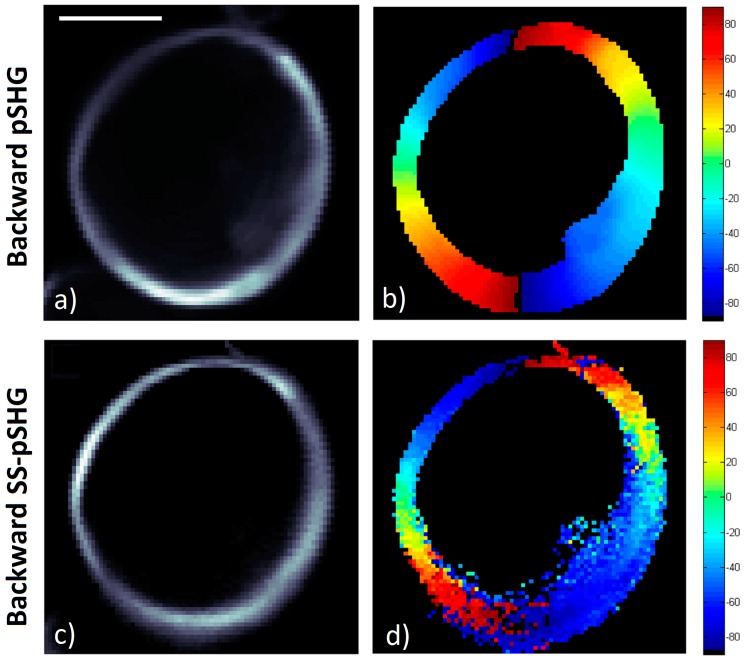
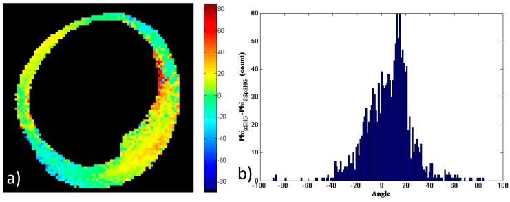
Polarization sensitive second harmonic generation (pSHG) microscopy is an imaging technique able to provide, in a non-invasive manner, information related to the molecular structure of second harmonic generation (SHG) active structures, many of which are commonly found in biological tissue. The process of acquiring this information by means of pSHG microscopy requires a scan of the sample using different polarizations of the excitation beam. This process can take considerable time in comparison with the dynamics of in vivo processes. Fortunately, single scan polarization sensitive second harmonic generation (SS-pSHG) microscopy has also been reported, and is able to generate the same information at a faster speed compared to pSHG. In this paper, the orientation of second harmonic active supramolecular assemblies in starch granules is obtained on by means of pSHG and SS-pSHG. These results are compared in the forward and backward directions, showing a good agreement in both techniques. This paper shows for the first time, to the best of the authors' knowledge, data acquired using both techniques over the exact same sample and image plane, so that they can be compared pixel-to-pixel.
Keywords: Medical and biological imaging; Nonlinear Microscopy; Polarization; Second harmonic generation.
Conflict of interest statement
The authors declare no conflict of interest.
Figures
Similar articles
-
Fast monitoring of in-vivo conformational changes in myosin using single scan polarization-SHG microscopy.Biomed Opt Express. 2014 Nov 24;5(12):4362-73. doi: 10.1364/BOE.5.004362. eCollection 2014 Dec 1. Biomed Opt Express. 2014. PMID: 25574444 Free PMC article.
-
Effect of molecular organization on the image histograms of polarization SHG microscopy.Biomed Opt Express. 2012 Oct 1;3(10):2681-93. doi: 10.1364/BOE.3.002681. Epub 2012 Sep 28. Biomed Opt Express. 2012. PMID: 23082306 Free PMC article.
-
Fast image analysis in polarization SHG microscopy.Opt Express. 2010 Aug 2;18(16):17209-19. doi: 10.1364/OE.18.017209. Opt Express. 2010. PMID: 20721110
-
Differential polarization nonlinear optical microscopy with adaptive optics controlled multiplexed beams.Int J Mol Sci. 2013 Sep 9;14(9):18520-34. doi: 10.3390/ijms140918520. Int J Mol Sci. 2013. PMID: 24022688 Free PMC article. Review.
-
Protein conformation and molecular order probed by second-harmonic-generation microscopy.J Biomed Opt. 2012 Jun;17(6):060901. doi: 10.1117/1.JBO.17.6.060901. J Biomed Opt. 2012. PMID: 22734730 Review.
Cited by
-
Second harmonic generation imaging reveals entanglement of collagen fibers in the elephant trunk skin dermis.Commun Biol. 2025 Jan 8;8(1):17. doi: 10.1038/s42003-024-07386-w. Commun Biol. 2025. PMID: 39779804 Free PMC article.
-
Double Stokes polarimetric microscopy for chiral fibrillar aggregates.Sci Rep. 2025 Feb 6;15(1):4464. doi: 10.1038/s41598-025-86893-0. Sci Rep. 2025. PMID: 39915558 Free PMC article.
-
Multiphoton microscopy is a nondestructive label-free approach to investigate the 3D structure of gas cell walls in bread dough.Sci Rep. 2023 Aug 26;13(1):13971. doi: 10.1038/s41598-023-39797-w. Sci Rep. 2023. PMID: 37634004 Free PMC article.
-
Polarimetric second-harmonic generation microscopy of partially oriented fibers I: Digital modeling.Biophys J. 2023 Oct 3;122(19):3924-3936. doi: 10.1016/j.bpj.2023.08.016. Epub 2023 Aug 22. Biophys J. 2023. PMID: 37608550 Free PMC article.
-
Polarimetric second harmonic generation microscopy of partially oriented fibers II: Imaging study.Biophys J. 2023 Oct 3;122(19):3937-3949. doi: 10.1016/j.bpj.2023.08.015. Epub 2023 Aug 23. Biophys J. 2023. PMID: 37621088 Free PMC article.
References
-
- Psilodimitrakopoulos S., Santos S.I., Amat-Roldan I., Thayil A.K., Artigas D., Loza-Alvarez P. In vivo, pixel-resolution mapping of thick filaments’ orientation in nonfibrilar muscle using polarization-sensitive second harmonic generation microscopy. J. Biomed. Opt. 2008;14:014001. doi: 10.1117/1.3059627. - DOI - PubMed
Grants and funding
LinkOut - more resources
Full Text Sources
